
Your Partner in Lifelong Health
Open Emergency Modal
Cutting-edge minimal access surgery ensu...
From joint replacements to complex traum...
Vice Chairman and Head
Minimal Access, Robotic & Bariatric Surgery
Vice Chairman
Medical Oncology
Vice-Chairman & Head
Radiation Oncology
Principal Director & Head
Pediatric Surgery & Pediatric Urology, Yashoda Cradle & Children's Hospital
Principal Director & Head
Department of Internal Medicine
Principal Director & Head
Obstetrics and Gynecology
Principal Director & Head
Gastroenterology and Hepatology
Principal Director & Unit Head
Neurosurgery
Senior Director and Head
Gynaecologic Oncology & Robotic Surgery
Senior Director
Urology, Uro-Oncology, Robotics & Renal Transplant
Director & Head
Pediatrics
Director & Unit Head
Orthopedics, Joint Replacement & Sports Medicine
Director and Unit Head
Cardio Thoracic and Vascular Surgery
Director
Cardiology
Director
Medical Oncology
Director
Orthopedics, Joint Replacement & Sports Medicine
Director
Yashoda Institute of Respiratory Medicine and Interventional Pulmonology
Associate Director
Internal Medicine
We offer you compassionate expert care through meticulous consultations, diagnostics, pharmacy and imaging, all of which together work to provide comprehensive and quality healthcare, all under one roof.
View All +
Care Beyond Boundaries
Stay informed with the latest announcements, medical breakthroughs, and hospital highlights.
Recognized by leading newspapers and media outlets for our excellence in healthcare.
From Critical Congenital Anomalies to a Thriving Newborn: A Multidisciplinary Neonatal Triumph
The journey of a preterm infant born with multiple life-threatening congenital anomalies represents one of the greatest challenges in modern neonatal medicine. This case highlights the successful management and remarkable recovery of a late preterm baby girl born at 34 weeks of gestation. Admitted with a complex clinical presentation—including Tracheo-Esophageal Fistula (TEF), respiratory distress, a right-sided rib fracture, and a Hemodynamically Significant Patent Ductus Arteriosus (Hs-PDA)—the newborn's rec...
From Critical Congenital Anomalies to a Thriving Newborn: A Multidisciplinary Neonatal Triumph
The journey of a preterm infant born with multiple life-threatening congenital anomalies represents one of the greatest challenges in modern neonatal medicine. This case highlights the successful management and remarkable recovery of a late preterm baby girl born at 34 weeks of gestation. Admitted with a complex clinical presentation—including Tracheo-Esophageal Fistula (TEF), respiratory distress, a right-sided rib fracture, and a Hemodynamically Significant Patent Ductus Arteriosus (Hs-PDA)—the newborn's recovery stands as a testament to advanced neonatal expertise and seamless multidisciplinary care. Through early diagnosis, timely surgical intervention, advanced intensive care ventilation and meticulous medical management, the infant progressed from a critical condition to becoming clinically stable, feeding orally and gaining weight. Today, her recovery reflects the world-class standard of care delivered by the Neonatal Intensive Care Unit (NICU) at Yashoda Medicity.
The patient, a late preterm female infant, was delivered via Lower Segment Caesarean Section (LSCS) due to maternal polyhydramnios and leaking amniotic fluid. Although she cried immediately after birth following stimulation, routine delivery room assessment revealed an inability to pass a feeding tube into the stomach. An urgent X-ray demonstrated coiling of the feeding tube within the upper oesophageal pouch, confirming the diagnosis of Tracheo-Esophageal Fistula (TEF). This congenital condition, characterised by an abnormal connection between the oesophagus and trachea, carries an immediate risk of aspiration, respiratory failure and inability to feed.
The infant soon developed severe respiratory distress requiring immediate Continuous Positive Airway Pressure (CPAP) support and subsequently mechanical ventilation following surgery. After extubation on the second post-operative day, she developed mild tachypnoea, tachycardia and a continuous cardiac murmur. A repeat echocardiogram confirmed a large Hemodynamically Significant Patent Ductus Arteriosus (Hs-PDA) with a left-to-right shunt, placing significant strain on her cardiovascular system. Managing these multiple complex conditions required close collaboration between the departments of Neonatology, Paediatric Surgery, Paediatric Cardiology and Orthopaedics.
The cornerstone of her recovery was the successful primary surgical repair of the Tracheo-Esophageal Fistula, performed on the fourth day of life by eminent Paediatric Surgeon Dr. Minu Bajpai. Following surgery, the baby received advanced mechanical ventilation using Synchronized Intermittent Mandatory Ventilation (SIMV) with Pressure Support and Volume Guarantee (VG) mode under sedation for five days. This lung-protective ventilation strategy effectively stabilised her respiratory status, allowing safe extubation to Non-Invasive Positive Pressure Ventilation (NIPPV) and gradual weaning to room air by the eighth post-operative day.
The development of a symptomatic Hemodynamically Significant Patent Ductus Arteriosus (Hs-PDA) following extubation required prompt medical management. Simultaneously, stringent infection prevention protocols were maintained throughout the hospital stay. Broad-spectrum intravenous antibiotics were administered empirically for eight days, and the infant remained completely free from sepsis during her admission.
Through meticulous round-the-clock neonatal monitoring and comprehensive multidisciplinary care, the baby achieved an excellent recovery. Her weight increased steadily from 2.168 kg at birth to 2.260 kg at discharge, demonstrating appropriate nutritional progress. On the eighth post-operative day, a fluoroscopic oesophagogram confirmed completely normal swallowing with no evidence of an anastomotic leak, allowing gradual advancement of feeds. She successfully transitioned from orogastric tube feeding to paladai feeds on day nine, established full oral feeding by day fourteen, and was breastfeeding on demand before discharge. At the time of discharge, the infant was neurologically normal, haemodynamically stable, feeding well and had a healthy surgical wound.
A key contributor to this successful outcome was Dr. Minu Bajpai, whose exceptional surgical expertise enabled precise anatomical reconstruction of the oesophagus in this fragile newborn. Her surgical excellence laid the foundation for the infant's complete recovery and long-term well-being.
This remarkable case demonstrates the transformative impact of modern neonatal medicine, where rapid diagnosis, advanced surgical expertise, sophisticated intensive care and coordinated multidisciplinary management can successfully overcome even the most complex congenital conditions, giving vulnerable newborns the opportunity to thrive and lead healthy lives.
Treatment By:
Dr. Minu Bajpai, Dr. Srijan Singh
Overcoming the Odds: A Neonatal Breakthrough in Severe RDS and Life-Threatening PPHN
In a definitive showcase of advanced critical care capabilities, Yashoda Medicity successfully managed and treated a highly volatile clinical crisis involving an early-term neonate suffering from severe Respiratory Distress Syndrome (RDS) and Persistent Pulmonary Hypertension of the Newborn (PPHN).
The male infant was delivered via Lower Segment Caesarean Section (LSCS) at 37 weeks of gestation and was immediately transferred to the Neonatal Intensive Care Unit (NICU) after developing early signs of respiratory distre...
Overcoming the Odds: A Neonatal Breakthrough in Severe RDS and Life-Threatening PPHN
In a definitive showcase of advanced critical care capabilities, Yashoda Medicity successfully managed and treated a highly volatile clinical crisis involving an early-term neonate suffering from severe Respiratory Distress Syndrome (RDS) and Persistent Pulmonary Hypertension of the Newborn (PPHN).
The male infant was delivered via Lower Segment Caesarean Section (LSCS) at 37 weeks of gestation and was immediately transferred to the Neonatal Intensive Care Unit (NICU) after developing early signs of respiratory distress, including marked nasal flaring, tachypnoea, audible grunting and significant chest wall retractions. On admission, the baby had a Downes score of 4/10, requiring urgent Non-Invasive Positive Pressure Ventilation (NIPPV) with a high oxygen requirement. Initial chest radiographs revealed low-volume lungs, confirming Respiratory Distress Syndrome (RDS) and necessitating immediate advanced neonatal respiratory support.
The infant's condition rapidly became more complex as respiratory distress worsened despite aggressive non-invasive ventilation. The neonatal team promptly administered the first dose of Curosurf® using the advanced Less Invasive Surfactant Administration (LISA) technique, resulting in temporary stabilisation. However, approximately 12 hours later, the baby's condition deteriorated significantly, with oxygen requirements increasing to 50% along with persistent respiratory retractions.
An urgent bedside 2D echocardiography, performed by Dr. Gaurav Kumar, Paediatric Cardiologist, revealed severe pulmonary arterial hypertension (PAH) with a dilated right atrium and right ventricle, a moderate-sized Patent Ductus Arteriosus (PDA) and an aneurysmal interatrial septum. At the same time, a repeat chest X-ray demonstrated progression to Grade 3 Respiratory Distress Syndrome, indicating worsening lung disease complicated by secondary Persistent Pulmonary Hypertension of the Newborn (PPHN).
Faced with these life-threatening complications, the multidisciplinary team immediately intubated the neonate, initiated Volume-Guaranteed (VG) mechanical ventilation, provided targeted sedation and administered a second dose of surfactant.
The success of this delicate recovery was led by the meticulous clinical expertise of Dr. Srijan Singh, Consultant Neonatologist, whose precise management of neonatal haemodynamics and advanced ventilator strategies proved lifesaving. Following the second dose of surfactant, the infant showed remarkable improvement, with oxygen requirements falling to 30% and arterial blood gas parameters returning to normal.
A follow-up echocardiogram performed within 24 hours confirmed complete resolution of severe pulmonary hypertension. This significant improvement allowed successful extubation back to NIPPV, followed by gradual weaning to Continuous Positive Airway Pressure (CPAP) over the next 48 hours. The infant was subsequently transitioned off all respiratory support and maintained stable spontaneous breathing.
By the eighth day of life, the newborn had made a complete recovery from severe Respiratory Distress Syndrome (RDS) and Persistent Pulmonary Hypertension of the Newborn (PPHN). The baby demonstrated normal neurological status, steady weight gain and complete haemodynamic stability, and was discharged in excellent health.
This case highlights the exceptional standard of neonatal intensive care at Yashoda Medicity, demonstrating how early diagnosis, timely surfactant therapy, advanced ventilatory support and coordinated multidisciplinary care can successfully manage even the most complex neonatal emergencies, leading to outstanding clinical outcomes.
Treatment By:
Dr. Srijan Singh, Dr. Gaurav Kumar
Rare Anterior Urethral Diverticulum Unmasked After Posterior Urethral Valve Ablation in a Male Infant
Posterior urethral valves (PUV) represent the most common cause of congenital bladder outlet obstruction in male infants(1,2). In contrast, anterior urethral valves (AUV) and associated congenital anterior urethral diverticula (CAUD) are rare, occurring 15–30 times less frequently(3). The coexistence of both proximal (PUV) and distal (AUV/CAUD) obstructions poses unique diagnostic challenges because the dominant proximal lesion can limit distension and mask the distal anomaly until after ablation(4). Lit...
Rare Anterior Urethral Diverticulum Unmasked After Posterior Urethral Valve Ablation in a Male Infant
Posterior urethral valves (PUV) represent the most common cause of congenital bladder outlet obstruction in male infants(1,2). In contrast, anterior urethral valves (AUV) and associated congenital anterior urethral diverticula (CAUD) are rare, occurring 15–30 times less frequently(3). The coexistence of both proximal (PUV) and distal (AUV/CAUD) obstructions poses unique diagnostic challenges because the dominant proximal lesion can limit distension and mask the distal anomaly until after ablation(4). Literature indicates that such combined anomalies may present with persistent or recurrent obstructive symptoms, urinary tract infections (UTIs), and upper tract deterioration post-PUV management(4–6). Early recognition via imaging (e.g., MCU) and cystoscopy is critical to prevent complications such as progressive renal damage, bladder dysfunction, or recurrent infections. This report describes a case of an anterior urethral diverticulum unmasked after PUV ablation, successfully managed with surgical reconstruction.
Case Report:
A 2-month-old male infant, born at term via normal vaginal delivery with unremarkableantenatal history, was referred as a follow-up case of PUV. On day 6 of life, he underwent cystoscopic ablation of PUV at our center for obstructive uropathy. Preoperative MCU on day2 of life demonstrated classic PUV findings . He presented at 2 months with high-grade fever (up to 102°F), poor urinary stream, dribbling, intermittent penile swelling that reduced after micturition, and dysuria. No hematuria, constipation, or neurological deficits were reported. Family history was unremarkable. On examination, the infant was irritable with mild dehydration. Abdominal palpation revealed a distended bladder. Genital examination showed ventral penile swelling that partially decompressed post-voiding . No hypospadias or other anomalies were noted. Vital signs were stable aside from fever. Labs showed leukocytosis, elevated C-reactive protein, and urine culture positive for Klebsiella pneumoniae (sensitive to targeted antibiotics). Mild azotemia was present. Ultrasonography confirmed bilateral
hydroureteronephrosis and thickened bladder wall.
After stabilization with antibiotics and bladder drainage, a repeat MCU demonstrated a
saccular outpouching consistent with an anterior urethral diverticulum/valve, delayed
emptying, residual dilated posterior urethra, trabeculated bladder, and bilateral grade III–IV
vesicoureteral reflux (VUR) or hydroureteronephrosis . The distal
anomaly had been masked by the prior proximal obstruction.
Following resolution of infection, the infant underwent cystoscopy and urethroplasty under
general anesthesia. Intraoperative findings confirmed a significant anterior urethral
diverticulum with a valve-like membrane. The base of the diverticulum was carefully incised
to create a controlled fistula for decompression of the obstructed system . This will
be followed by meticulous urethroplasty: excision of redundant diverticular tissue,reconstruction of the urethral continuity using fine absorbable sutures to ensure a wide caliber
lumen without tension, and multilayer closure to support the repair.
The patient remained stable perioperatively. He was monitored for 24 hours with analgesia
and prophylactic antibiotics. He was discharged on postoperative day 14, afebrile, feeding
well, gaining weight, and voiding with a good stream. Follow-up ultrasonography showed
improving hydroureteronephrosis. Long-term surveillance for renal function, bladder
dynamics, and stricture is planned.
Discussion:
The association of anterior urethral diverticulum/valve with PUV is rare, with limited cases
reported(7). The proximal PUV restricts distal urethral filling, often delaying manifestation of
the anterior lesion until after ablation(4). Persistent symptoms post-PUV treatment should
prompt evaluation for concomitant distal anomalies(7).
MCU is the gold standard for diagnosis, revealing the diverticular outpouching, “spinning
top” urethra, and associated VUR or hydronephrosis(8,9). Cystoscopy confirms the valve-like
membrane and diverticulum(7).
Management is individualized based on size and tissue support. Small valves may respond to
endoscopic fulguration or laser ablation. Larger saccular diverticula, as in this case, require
open diverticulectomy and urethroplasty to excise redundant tissue, reconstruct continuity,
reduce dead space, and prevent complications (e.g., stones, recurrent infection, or
stricture)(3,10). Techniques range from primary anastomosis to staged repairs or substitution
urethroplasty in complex cases(11–13).
This case achieved excellent short-term outcomes with controlled decompression and
meticulous reconstruction. Potential complications of untreated CAUD include renal
deterioration and bladder decompensation(14,15). Thorough endoscopic evaluation and
individualized surgery are essential.
Treatment By:
Dr. Minu Bajpai, Dr. Srijan Singh
Complete Duodenal Obstruction Due to Transmesenteric Internal Bowel Herniation and Congenital Bands: A Lifesaving Neonatal Intervention
In a remarkable clinical milestone that underscores the power of swift, multidisciplinary intervention, Yashoda Medicity successfully managed and treated a highly critical, life-threatening congenital anomaly in a 4-day-old neonate. The male infant, Baby of Pushpa, was admitted to the Neonatal Intensive Care Unit (NICU) following a critical referral, presenting with an acute history of multiple episodes of bilious vomiting, significant abdominal distension, and sluggish bow...
Complete Duodenal Obstruction Due to Transmesenteric Internal Bowel Herniation and Congenital Bands: A Lifesaving Neonatal Intervention
In a remarkable clinical milestone that underscores the power of swift, multidisciplinary intervention, Yashoda Medicity successfully managed and treated a highly critical, life-threatening congenital anomaly in a 4-day-old neonate. The male infant, Baby of Pushpa, was admitted to the Neonatal Intensive Care Unit (NICU) following a critical referral, presenting with an acute history of multiple episodes of bilious vomiting, significant abdominal distension, and sluggish bowel sounds. Recognizing the extreme urgency of the situation, the clinical team immediately initiated stabilization protocols, making the baby nil per os (NPO), commencing intravenous fluid resuscitation, administering empirical antibiotics, and inserting a nasogastric/orogastric tube. Initial abdominal radiographs revealed a severely dilated stomach paired with a concerning gasless abdomen, signaling a high-grade gastrointestinal obstruction that required immediate expert diagnostic elucidation and surgical evaluation.
The case rapidly transformed into a collaborative triumph through the immediate involvement of the hospital's elite pediatric medical leadership. Dr. Minu Bajpai, an eminent and veteran pediatric surgeon whose vast experience and decisive clinical acumen are widely recognized, was promptly consulted to guide the surgical trajectory. Dr. Bajpai recommended a gastrograffin contrast study, which definitively demonstrated a complete mechanical obstruction at the level of the second part of the duodenum alongside proximal gastro-duodenal dilatation. Armed with this critical insight, Dr. Bajpai led the surgical team in performing an emergent exploratory laparotomy. The intraoperative findings revealed a rare and treacherous internal internal herniation of the small bowel through the transverse mesocolon, compounded by restrictive congenital bands compromising the second part of the duodenum. With meticulous precision, the surgical team divided the bands and successfully achieved complete deherniation of the entrapped bowel, resolving the primary anatomical crisis.
Following the intricate surgical procedure, the complex task of critical post-operative management fell to the highly accomplished neonatologist, Dr. Srijan Singh, whose specialized dedication to neonatal intensive care proved pivotal to the infant’s survival. Dr. Singh orchestrated a comprehensive, advanced recovery plan in the NICU, keeping the fragile neonate NPO and introducing Total Parenteral Nutrition (TPN) via a peripherally inserted central catheter (PICC) line to sustain metabolic demands. Under Dr. Singh’s vigilant oversight, the baby received tailored mechanical ventilation for 36 hours before being successfully extubated to room air. Dr. Singh ordered a repeat gastrograffin study, which confirmed a perfectly patent, non-obstructed post-operative duodenum with excellent contrast passage.
The synergistic collaboration between the surgical prowess of Dr. Minu Bajpai and the meticulous, expert neonatal care of Dr. Srijan Singh ensured a flawless trajectory toward recovery. Over the subsequent days, enteral feeds were gradually advanced, the post-operative sepsis screen remained entirely negative, and blood cultures proved sterile, allowing the safe discontinuation of intravenous antibiotics. By the tenth post-operative day, the infant demonstrated a complete clinical turnaround—actively accepting full breastfeeds and paladai feeds, passing normal stools without any vomiting, and steadily gaining weight. The infant was discharged in a hemodynamically stable condition with a healthy, healing surgical site. This extraordinary breakthrough highlights the world-class neonatal and pediatric surgical capabilities available at Yashoda Medicity, showcasing how seamless teamwork, clinical expertise, and cutting-edge intensive care can rewrite the medical outcomes for the most vulnerable patients.
Treatment By:
Dr. Minu Bajpai, Dr. Srijan Singh
A primigravida mother with pregnancy-induced hypertension (PIH) and deranged antenatal Dopplers (reversal of flow in ductus venosus) delivered baby via emergency LSCS at 30 weeks. Baby had extremely low birth weight (900 grams) and had been diagnosed with severe fetal growth restriction (FGR) antenatally and presented with perinatal depression and respiratory distress immediately after birth.
Neonatal Course & Challenges
This extremely preterm, extremely low birth weight (ELBW) infant faced several serious complications typical of extreme prematurity:
A primigravida mother with pregnancy-induced hypertension (PIH) and deranged antenatal Dopplers (reversal of flow in ductus venosus) delivered baby via emergency LSCS at 30 weeks. Baby had extremely low birth weight (900 grams) and had been diagnosed with severe fetal growth restriction (FGR) antenatally and presented with perinatal depression and respiratory distress immediately after birth.
Neonatal Course & Challenges
This extremely preterm, extremely low birth weight (ELBW) infant faced several serious complications typical of extreme prematurity:
Despite these multiple challenges, the baby showed remarkable resilience.
Multidisciplinary Management
Under the leadership of Dr. Srijan Singh, the NICU team at Yashoda Medicity provided comprehensive, evidence-based care:
The baby remained hemodynamically stable in the later part of the stay, achieved full oral (paladai) feeds, and maintained temperature and vitals without support.
Baby was neurologically normal at discharge and gaining weight. Baby did not have any ROP, BPD or IVH.
Treatment By:
Dr. Srijan Singh, Dr. Samreen Zehra
Advanced neonatal care plays a critical role in improving survival and long-term outcomes for extremely preterm infants. In a recent case, our multidisciplinary team successfully managed the delivery and care of a baby boy born at just 26 weeks of gestation, weighing under 800 grams.
The newborn required immediate respiratory support and was admitted to a Level III Neonatal Intensive Care Unit (NICU), where he received round-the-clock monitoring and specialised care. Through meticulous medical management and dedicated nursing support, the baby demonstrated consistent progress over the course of treatment.
He was d...
Advanced neonatal care plays a critical role in improving survival and long-term outcomes for extremely preterm infants. In a recent case, our multidisciplinary team successfully managed the delivery and care of a baby boy born at just 26 weeks of gestation, weighing under 800 grams.
The newborn required immediate respiratory support and was admitted to a Level III Neonatal Intensive Care Unit (NICU), where he received round-the-clock monitoring and specialised care. Through meticulous medical management and dedicated nursing support, the baby demonstrated consistent progress over the course of treatment.
He was discharged in a stable condition, breathing independently and feeding well, marking a significant milestone in his recovery journey.
This case was led by Dr. Neha Agrawal, Consultant – Pediatrics & Neonatology; Dr. J.B. Sharma, Principal Director & Head – Obstetrics and Gynaecology; Dr. Namita Nigam Datta, Senior Consultant – Obstetrics and Gynaecology; and Dr. Nisha, Consultant – Obstetrics & Gynaecology, alongside our experienced nursing and clinical teams.
It reflects the strength of coordinated clinical expertise, advanced neonatal infrastructure, and a commitment to delivering specialised care focused on giving every newborn the best possible start in life.
Treatment By:
Dr. Neha Agrawal, Dr. J.B. Sharma, Dr. Namita Nigam Datta, Dr. Nisha
A 7-year-old girl presented with persistent pain in the right knee for two months. Initial investigations, including X-ray and MRI, raised suspicion of a malignant lesion in the distal femur near the knee joint.
A biopsy confirmed the diagnosis of osteosarcoma, a rare bone malignancy seen in children and adolescents. A metastatic workup was conducted to assess disease spread, which showed no evidence of metastasis.
The patient was initiated on a structured treatment protocol involving multiple cycles of neoadjuvant chemotherapy, followed by planned surgical intervention and postoperative chemotherapy.
After comp...
A 7-year-old girl presented with persistent pain in the right knee for two months. Initial investigations, including X-ray and MRI, raised suspicion of a malignant lesion in the distal femur near the knee joint.
A biopsy confirmed the diagnosis of osteosarcoma, a rare bone malignancy seen in children and adolescents. A metastatic workup was conducted to assess disease spread, which showed no evidence of metastasis.
The patient was initiated on a structured treatment protocol involving multiple cycles of neoadjuvant chemotherapy, followed by planned surgical intervention and postoperative chemotherapy.
After completion of four chemotherapy cycles, repeat imaging demonstrated a favorable response, and surgical management was undertaken. Considering the child’s age and significant remaining growth potential, the key challenge was to achieve oncological clearance while preserving future limb growth.
A customised expandable prosthesis was designed for the patient to accommodate longitudinal bone growth through future length adjustments. The surgical procedure was completed successfully, and the postoperative course remained stable and uneventful.
The patient continues under follow-up, with the focus on long-term recovery, functional preservation, and support for normal growth and development.
Treatment By:
Dr. Nikhil Tandon
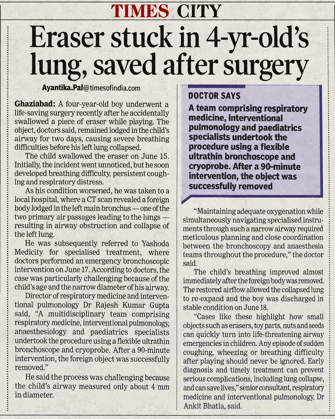
Advanced Bronchoscopic Intervention at Yashoda Medicity Removes Eraser from 4-Year-Old's Airway, Restores Collapsed Lung.
Doctors at Yashoda Medicity successfully saved a 4-year-old child after a piece of eraser became lodged in his left main bronchus, causing near-complete airway obstruction and collapse of the left lung. The 4-year old child accidentally inhaled the eraser while playing nearly two days before reaching the hospital.
An emergency bronchoscopic procedure was performed using a flexible ultrathin bronchoscope and cryoprobe. The multidisciplinary team included Dr Rajesh Kumar Gupta, Dire...
Advanced Bronchoscopic Intervention at Yashoda Medicity Removes Eraser from 4-Year-Old's Airway, Restores Collapsed Lung.
Doctors at Yashoda Medicity successfully saved a 4-year-old child after a piece of eraser became lodged in his left main bronchus, causing near-complete airway obstruction and collapse of the left lung. The 4-year old child accidentally inhaled the eraser while playing nearly two days before reaching the hospital.
An emergency bronchoscopic procedure was performed using a flexible ultrathin bronchoscope and cryoprobe. The multidisciplinary team included Dr Rajesh Kumar Gupta, Director; Dr Ankit Bhatia, Senior Consultant in Respiratory Medicine and Interventional Pulmonology; Dr Lokesh, Principal Director & HOD of Anesthesiology & Critical Care; and Dr (Maj) Shaifali Bhatia, Senior Consultant - Paediatrics & Neonatology.
During the 90-minute procedure, doctors successfully removed the eraser and restored airflow to the collapsed lung. The intervention was particularly challenging because the child's airway measured only about 4 mm in diameter, requiring precise airway management and close coordination between the bronchoscopy and anaesthesia teams.
Speaking about the case, Dr Rajesh Kumar Gupta, Director, Respiratory Medicine and Interventional Pulmonology, Yashoda Medicity, said, “This was a particularly challenging procedure because the child’s airway measured only about 4 mm in diameter. Maintaining adequate oxygenation while simultaneously navigating specialized instruments through such a narrow airway required meticulous planning and close coordination between the bronchoscopy and anaesthesia teams over a 90-minute procedure.
Dr Ankit Bhatia, Senior Consultant, Respiratory Medicine & Interventional Pulmonology, said, “Cases like these highlight how small objects such as erasers, toy parts, nuts, and seeds can quickly turn into life-threatening airway emergencies in children. Any episode of sudden coughing, wheezing, or breathing difficulty after playing should never be ignored. Early diagnosis and timely treatment can prevent serious complications, including lung collapse, and can save lives.”
Describing the ordeal as one of the most frightening experiences for the family, the child's parent said, “Watching him struggle to breathe was one of the most frightening experiences for our family. We are deeply grateful to the doctors and medical team at Yashoda Medicity for their timely intervention, expertise, and constant support throughout the treatment. Knowing that specialists from different disciplines were working together and that advanced facilities were available gave us immense confidence during a very difficult time. We hope other parents learn from our experience and seek immediate medical attention if a child develops persistent coughing, wheezing, or breathing difficulty after inhaling or choking on a small object.”
Following removal of the foreign body, the child's breathing improved immediately. Airflow to the left lung was restored, allowing the collapsed lung to re-expand. The child recovered well and was discharged in stable condition on 18 June 2026.
Doctors emphasized that young children are particularly vulnerable to accidentally inhaling small objects during play. In this case, the foreign body had already caused the collapse of the left lung. Further delay in treatment could have resulted in severe infection, persistent lung collapse, irreversible lung damage, respiratory failure, and potentially life-threatening complications.
The successful outcome underscores the importance of timely diagnosis, advanced bronchoscopic expertise, specialized equipment, and a multidisciplinary approach in managing complex airway emergencies in children. Yashoda Medicity remains committed to providing comprehensive, advanced interventional pulmonology and critical care services, backed by state-of-the-art technology and specialist teams capable of handling complex medical emergencies across age groups.
Treatment By:
Dr Rajesh Kumar Gupta, Director; Dr Ankit Bhatia, Dr Lokesh; and Dr (Maj) Shaifali Bhatia.
A G6A5L0 mother with an IVF-conceived pregnancy had a history of threatened abortion and intrauterine demise of Twin A at 22 weeks of gestation.
Twin B, a 24-week gestation male/female preterm neonate weighing 650 g, was delivered by emergency LSCS in view of placental abruption. The baby cried immediately after birth but developed severe respiratory distress and was promptly shifted to the Level III NICU for intensive care.
The neonate received three doses of surfactant therapy and required invasive mechanical ventilation for 7 days, followed by non-invasive respiratory support, with gradual weaning to room air...
A G6A5L0 mother with an IVF-conceived pregnancy had a history of threatened abortion and intrauterine demise of Twin A at 22 weeks of gestation.
Twin B, a 24-week gestation male/female preterm neonate weighing 650 g, was delivered by emergency LSCS in view of placental abruption. The baby cried immediately after birth but developed severe respiratory distress and was promptly shifted to the Level III NICU for intensive care.
The neonate received three doses of surfactant therapy and required invasive mechanical ventilation for 7 days, followed by non-invasive respiratory support, with gradual weaning to room air. Enteral feeding was initially limited due to feed intolerance, and the baby remained on total parenteral nutrition (TPN) until full enteral feeds were successfully established by Day 18 of life.
During the NICU stay, the baby was treated with intravenous antibiotics for 2 weeks for sepsis. Subsequently, the baby developed fungal sepsis, which was successfully managed with appropriate antifungal therapy.
Serial cranial ultrasonography was performed as per protocol and showed no evidence of intraventricular hemorrhage (IVH) or periventricular leukomalacia (PVL).
ROP screening was initially normal. However, at 33 weeks postmenstrual age, the baby developed pre-plus disease in Zone II, which required laser photocoagulation therapy. Subsequent follow-up examinations documented satisfactory regression of ROP.
The baby also required packed red blood cell (PRBC) transfusion during the hospital stay for anemia of prematurity.
The case was successfully managed by a multidisciplinary team comprising Dr. Minu Bajpai, Dr. J.B. Sharma, Dr. Neha, Dr. Samreen, Dr. Divya Arora, Dr. Ayush Jain, and Dr. Nisha.
After a prolonged NICU stay of 78 days, the baby was discharged in a clinically stable condition on room air, tolerating spoon feeds and breastfeeding well, with a discharge weight of 1,625 g.
Treatment By:
Dr Neha Agrawal (Consultant Neonatologist ) and NICU team.
Robotic Milestone in Advanced Cancer Surgery: Combined Robotic LAR with Portal Vein Ligation
Our surgical oncology team recently achieved another important milestone in minimally invasive cancer surgery by successfully performing a Robotic Low Anterior Resection (LAR) combined with robotic ligation of the right portal vein in a 50-year-old female diagnosed with rectal cancer and synchronous liver metastasis.
The patient presented with rectal adenocarcinoma along with metastatic disease involving the right lobe of the liver. Managing such complex cases requires meticulous planning to achieve complete o...
Robotic Milestone in Advanced Cancer Surgery: Combined Robotic LAR with Portal Vein Ligation
Our surgical oncology team recently achieved another important milestone in minimally invasive cancer surgery by successfully performing a Robotic Low Anterior Resection (LAR) combined with robotic ligation of the right portal vein in a 50-year-old female diagnosed with rectal cancer and synchronous liver metastasis.
The patient presented with rectal adenocarcinoma along with metastatic disease involving the right lobe of the liver. Managing such complex cases requires meticulous planning to achieve complete oncological clearance while preserving adequate liver function.
After multidisciplinary tumor board discussion, a staged surgical strategy was planned with curative intent. In the first stage, a robotic low anterior resection was performed following total mesorectal excision principles to remove the rectal primary tumor. During the same sitting, robotic ligation of the right branch of the portal vein was carried out. This step is designed to induce hypertrophy of the future liver remnant, allowing safe completion of a planned right hepatectomy after approximately four weeks.
The use of robotic technology enabled precise pelvic dissection, superior visualization, and delicate hilar vascular control, allowing two major oncologic objectives to be achieved through a minimally invasive approach. The patient tolerated the procedure well and is recovering satisfactorily while awaiting the second-stage liver surgery.
This combined robotic approach highlights the evolving role of advanced minimally invasive surgery in complex gastrointestinal and hepatobiliary oncology. By integrating colorectal and liver strategies in a single operation, patients benefit from reduced surgical trauma, faster recovery, and preservation of curative treatment pathways.
The case reflects our institution’s continued commitment to innovation, multidisciplinary collaboration, and delivery of world-class cancer care using cutting-edge robotic surgical techniques.
Treatment By:
Dr. Asim Rizvi
A 59-year-old female from Comoros presented with severe bilateral knee dysfunction and associated implant-related complications. She had a Grade 3 valgus deformity with advanced osteoarthritis, a failed primary total knee replacement of the left knee, and significant implant impingement at the left ankle, resulting in marked pain and restricted mobility.
She was evaluated by the orthopaedic team at Yashoda Medicity led by Dr. Brajesh Koushle and Dr. Gaurav Thakral, at Yashoda Medicity. Given the complexity of her condition and functional limitations, a comprehensive surgical plan was formulated.
The patient unde...
A 59-year-old female from Comoros presented with severe bilateral knee dysfunction and associated implant-related complications. She had a Grade 3 valgus deformity with advanced osteoarthritis, a failed primary total knee replacement of the left knee, and significant implant impingement at the left ankle, resulting in marked pain and restricted mobility.
She was evaluated by the orthopaedic team at Yashoda Medicity led by Dr. Brajesh Koushle and Dr. Gaurav Thakral, at Yashoda Medicity. Given the complexity of her condition and functional limitations, a comprehensive surgical plan was formulated.
The patient underwent primary total knee replacement of the right knee, revision total knee replacement of the left knee, and removal of the impinging implant from the left ankle in the same sitting. The surgery was successfully completed with meticulous intraoperative management.
Postoperatively, the patient showed excellent recovery. Early mobilisation was initiated, and she was able to stand and walk from the first post-operative day. She was discharged in a stable condition, walking unassisted and with significant improvement in functional outcomes.
Treatment By:
Dr. Brajesh Koushle, Dr. Gaurav Thakral, and Team
A 65-year-old female, a known carcinoma colon survivor, presented with altered sensorium, severe headache, diplopia, and worsening vision.
On arrival, she was drowsy, disoriented, with gaze palsy, hemianopia, and significantly elevated blood pressure. MRI brain revealed a large left occipital tumor with acute haemorrhage, requiring emergency intervention.
She underwent urgent craniotomy with total tumor excision and evacuation of the hematoma. The surgery was completed successfully.
Post-operatively, she showed rapid and remarkable recovery, with significant improvement in neurological status. She was discharge...
A 65-year-old female, a known carcinoma colon survivor, presented with altered sensorium, severe headache, diplopia, and worsening vision.
On arrival, she was drowsy, disoriented, with gaze palsy, hemianopia, and significantly elevated blood pressure. MRI brain revealed a large left occipital tumor with acute haemorrhage, requiring emergency intervention.
She underwent urgent craniotomy with total tumor excision and evacuation of the hematoma. The surgery was completed successfully.
Post-operatively, she showed rapid and remarkable recovery, with significant improvement in neurological status. She was discharged in four days and, at one-week follow-up, was walking independently with restored clarity of vision and no headache.
Her recovery was exceptional for the extent of pathology and age, with neurological improvement far exceeding expected timelines.
Treatment By:
Dr. Rajinder Kumar
Minimally invasive and Scarless solution for Brain aneurysm at Yashoda Medicity
A patient with a complex brain aneurysm was treated at Yashoda Medicity through a minimally invasive and scarless approach. This was the first neurointervention for a complex brain aneurysm in the state, performed in our state-of-the-art Philips Biplanar Cath Lab, equipped with advanced softwares including 3D angiography, CBCT, Xper CT, VasoCT, MAFA, and Clarity IQ.
The procedure was led by Dr. Dibya Jyoti Mahakul, Consultant – Endovascular Neurosurgery, who carefully placed a Flow Diverter Stent across the aneurysm. Us...
Minimally invasive and Scarless solution for Brain aneurysm at Yashoda Medicity
A patient with a complex brain aneurysm was treated at Yashoda Medicity through a minimally invasive and scarless approach. This was the first neurointervention for a complex brain aneurysm in the state, performed in our state-of-the-art Philips Biplanar Cath Lab, equipped with advanced softwares including 3D angiography, CBCT, Xper CT, VasoCT, MAFA, and Clarity IQ.
The procedure was led by Dr. Dibya Jyoti Mahakul, Consultant – Endovascular Neurosurgery, who carefully placed a Flow Diverter Stent across the aneurysm. Using MAFA software, the first of its kind in North India, the team successfully achieved and recorded the target aneurysmal flow parameters.
The use of these advanced technologies aided in faster decision-making, reduced radiation exposure, and improved procedural precision, reflecting our belief that “Time is Brain.” This case demonstrates our preparedness to manage all varieties of complex neurovascular conditions, from ischemic stroke to hemorrhagic stroke, through minimally invasive and scarless procedures.
When it comes to neurointervention and endovascular neurosurgery, Yashoda Medicity, equipped with the latest technologies and expert teams, is miles ahead in delivering advanced patient care.
Treatment By:
Dr. Dibya Jyoti Mahakul
A 27-year-old patient diagnosed with advanced-stage T-cell lymphoma presented for specialised evaluation and management. Given the aggressive nature of the disease, a detailed clinical assessment and a structured treatment plan were undertaken by the expert team.
The patient was treated with high-dose chemotherapy followed by an autologous stem cell transplant, a well-established approach for managing advanced hematological malignancies. The treatment was carried out under the expertise of Dr. Nivedita Dhingra, Director & Head – Hematology, Hemato-Oncology and Bone Marrow Transplant (Adult & Pediatrics), along wit...
A 27-year-old patient diagnosed with advanced-stage T-cell lymphoma presented for specialised evaluation and management. Given the aggressive nature of the disease, a detailed clinical assessment and a structured treatment plan were undertaken by the expert team.
The patient was treated with high-dose chemotherapy followed by an autologous stem cell transplant, a well-established approach for managing advanced hematological malignancies. The treatment was carried out under the expertise of Dr. Nivedita Dhingra, Director & Head – Hematology, Hemato-Oncology and Bone Marrow Transplant (Adult & Pediatrics), along with Dr. Karuna Jha, Senior Consultant – Hematology, Hemato-Oncology and Bone Marrow Transplant (Adult & Pediatrics), supported by the dedicated BMT team.
The treatment protocol was designed to achieve optimal disease control while ensuring patient safety. High-dose chemotherapy helped target and eliminate malignant cells, followed by stem cell reinfusion to restore healthy bone marrow function.
The patient responded well to the treatment and was closely monitored throughout the recovery phase, demonstrating a stable clinical course post-procedure.
This case highlights the importance of a protocol-based, multidisciplinary approach in managing rare and complex lymphomas, combining clinical expertise with advanced transplant capabilities to achieve effective outcomes.
Treatment By:
Dr. Nivedita Dhingra & Dr. Karuna Jha
A patient diagnosed with a tumour in the head of the pancreas presented for specialised evaluation and management. Given the complexity of the condition, a detailed clinical assessment and surgical planning were undertaken by the expert team.
The patient was planned for a Robotic Pancreaticoduodenectomy (Whipple’s Procedure), a highly specialised surgery used to treat tumours in this region. The procedure was performed under the expertise of Dr. Syed Asim Rizvi, Principal Director – Surgical Oncology and Robotic Surgery, along with his team, using advanced robotic surgical technology.
The robotic approach provided...
A patient diagnosed with a tumour in the head of the pancreas presented for specialised evaluation and management. Given the complexity of the condition, a detailed clinical assessment and surgical planning were undertaken by the expert team.
The patient was planned for a Robotic Pancreaticoduodenectomy (Whipple’s Procedure), a highly specialised surgery used to treat tumours in this region. The procedure was performed under the expertise of Dr. Syed Asim Rizvi, Principal Director – Surgical Oncology and Robotic Surgery, along with his team, using advanced robotic surgical technology.
The robotic approach provided enhanced precision and superior visualisation, enabling meticulous removal of the tumour while preserving surrounding structures. This minimally invasive technique also helped reduce intraoperative blood loss and supported a more controlled surgical environment.
The patient demonstrated a smooth postoperative recovery with reduced pain, early mobilisation, and overall improved comfort. He was discharged in stable condition.
This case highlights the role of advanced robotic surgery in managing complex pancreatic tumours, combining surgical expertise with precision technology to achieve optimal patient outcomes.
Treatment By:
Dr. Asim Rizvi
A 78-year-old male patient presented with difficulty in swallowing and a small growth at the base of the tongue. Initial evaluation, including biopsy and imaging, confirmed a malignancy involving the tongue along with an affected lymph node in the neck.
As part of routine staging, a whole-body scan revealed an additional lesion in the upper lobe of the lung with associated chest lymph nodes. Further evaluation confirmed this to be a second, unrelated primary cancer. This rare presentation of two simultaneous primary cancers required a carefully coordinated and highly specialised treatment approach.
After a detailed as...
A 78-year-old male patient presented with difficulty in swallowing and a small growth at the base of the tongue. Initial evaluation, including biopsy and imaging, confirmed a malignancy involving the tongue along with an affected lymph node in the neck.
As part of routine staging, a whole-body scan revealed an additional lesion in the upper lobe of the lung with associated chest lymph nodes. Further evaluation confirmed this to be a second, unrelated primary cancer. This rare presentation of two simultaneous primary cancers required a carefully coordinated and highly specialised treatment approach.
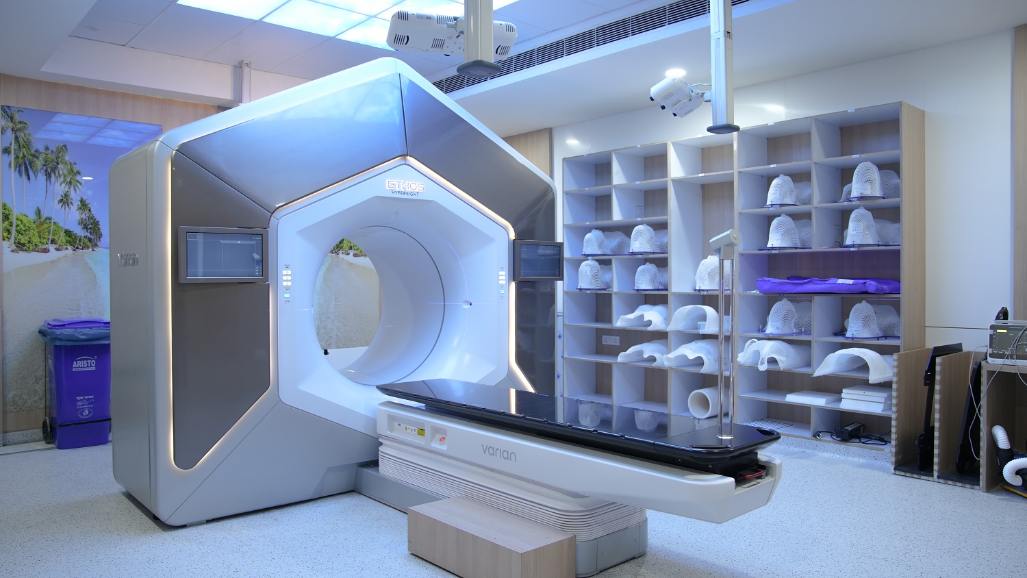
After a detailed assessment by a multidisciplinary team, a comprehensive treatment plan was developed with curative intent. The patient was treated using advanced adaptive radiotherapy, a technique that allows real-time adjustments to radiation delivery based on daily imaging and anatomical changes, ensuring high precision and safety.
Both cancers were treated concurrently. The lung and chest lymph nodes received a planned course of radiation over six weeks, while the tongue and neck region were treated over seven weeks. Each session was guided by high-resolution imaging, enabling accurate targeting of the tumours while preserving surrounding healthy structures. The patient completed the treatment successfully with good tolerance. He showed a steady recovery and continues to remain stable on follow-up.
This case highlights the power of advanced radiation technology and multidisciplinary expertise in managing rare and complex cancer presentations.
Treatment By:
Dr. Gagan Saini
A 55-year-old woman diagnosed with advanced high-grade serous ovarian cancer presented after completing three cycles of neoadjuvant chemotherapy. She had responded well to treatment, with imaging showing significant disease resolution and tumour markers returning to normal levels. Following a comprehensive evaluation, she was planned for interval debulking surgery combined with Heated Intraperitoneal Chemotherapy (HIPEC).
After detailed counselling and preoperative optimisation, the patient underwent surgery under the care of the expert team. A meticulous cytoreductive procedure was performed to remove all visible disease, ...
A 55-year-old woman diagnosed with advanced high-grade serous ovarian cancer presented after completing three cycles of neoadjuvant chemotherapy. She had responded well to treatment, with imaging showing significant disease resolution and tumour markers returning to normal levels. Following a comprehensive evaluation, she was planned for interval debulking surgery combined with Heated Intraperitoneal Chemotherapy (HIPEC).
After detailed counselling and preoperative optimisation, the patient underwent surgery under the care of the expert team. A meticulous cytoreductive procedure was performed to remove all visible disease, followed by the administration of HIPEC, a targeted chemotherapy delivered directly into the abdominal cavity using specialised equipment over a controlled duration. The entire procedure was carried out with precise monitoring and a highly coordinated approach.
Postoperatively, the patient was closely monitored with a structured care protocol, including optimal fluid management and regular assessment of clinical parameters to support recovery.
The patient demonstrated a smooth and steady recovery and was discharged in stable condition on the eighth postoperative day. She continues to do well on follow-up.
This case marks a significant milestone as one of the successful HIPEC procedures performed at Yashoda Medicity, highlighting the team’s expertise in delivering advanced oncological care with precision and safety.
Treatment By:
Dr. Satinder Kaur, Dr. Hemlata Garg, and Dr. Ipshita Sahoo
A 65-year-old male patient, weighing 44 kg, presented with long-standing coronary artery disease, a recent myocardial infarction, critical multivessel disease, unstable angina, and severe left ventricular dysfunction with a left ventricular ejection fraction (LVEF) of 20–25%. Considering the complexity of his condition, he underwent coronary artery bypass grafting (CABG) under the care of the expert cardiac team.
The procedure was successfully performed, with the patient requiring minimal inotropic support and no intra-aortic balloon pump (IABP). He was extubated within four hours, reflecting a stable and well-managed imm...
A 65-year-old male patient, weighing 44 kg, presented with long-standing coronary artery disease, a recent myocardial infarction, critical multivessel disease, unstable angina, and severe left ventricular dysfunction with a left ventricular ejection fraction (LVEF) of 20–25%. Considering the complexity of his condition, he underwent coronary artery bypass grafting (CABG) under the care of the expert cardiac team.
The procedure was successfully performed, with the patient requiring minimal inotropic support and no intra-aortic balloon pump (IABP). He was extubated within four hours, reflecting a stable and well-managed immediate postoperative course.
Post-surgery, the patient was closely monitored and managed with a highly calibrated clinical approach. Advanced hemodynamic support strategies, including the timely use of an intra-aortic balloon pump (IABP), along with optimised inotropic management, judicious fluid administration, and targeted medical therapy, ensured effective systemic and organ perfusion.
Over the next few days, the patient demonstrated steady clinical improvement, with stabilising metabolic parameters, improving organ function, and gradual return to normal physiological activity. This enabled the phased withdrawal of advanced support measures, including ventilatory and circulatory assistance.
Following a structured period of intensive care and rehabilitation, the patient was discharged in stable condition. The case highlights the team’s expertise in managing complex high-risk cardiac conditions with precision, resulting in a successful recovery.
Treatment By:
Dr. Rachit Saxena, Dr. Vikas Kumar Keshari, and Dr. Kushant Gupta
A 28-year-old male from Nepal was diagnosed with chronic kidney disease after experiencing persistent weakness and loss of appetite for nearly two years. Investigations revealed severely impaired kidney function, and he was initiated on hemodialysis through a catheter placed in the right side of his neck. He was advised to undergo AV fistula creation and maintain regular maintenance hemodialysis (MHD) three times a week.
However, due to irregular dialysis sessions, his condition worsened. He then consulted the expert team of nephrologists, Dr. Prajit Mazumdar and Dr. Inderjit G. Momin, along with urologists, Dr. Vaibhav Sax...
A 28-year-old male from Nepal was diagnosed with chronic kidney disease after experiencing persistent weakness and loss of appetite for nearly two years. Investigations revealed severely impaired kidney function, and he was initiated on hemodialysis through a catheter placed in the right side of his neck. He was advised to undergo AV fistula creation and maintain regular maintenance hemodialysis (MHD) three times a week.
However, due to irregular dialysis sessions, his condition worsened. He then consulted the expert team of nephrologists, Dr. Prajit Mazumdar and Dr. Inderjit G. Momin, along with urologists, Dr. Vaibhav Saxena and Dr. Kuldeep Agarwal, at Yashoda Medicity. The team counselled him regarding renal replacement therapy (RRT), the importance of regular MHD, and recommended renal transplantation as the most effective long-term solution.
His transplant evaluation began with his mother as the donor. During the assessment, he was found to have significant anaemia and reduced cardiac function, likely resulting from inconsistent dialysis, which increased his peri-operative risk. After thorough counselling, he was optimised for surgery, and immunosuppressive therapy was initiated two days prior to the procedure.
The patient successfully underwent renal transplantation with ATG induction. Post-surgery, he showed excellent recovery with good urine output and steadily improving creatinine levels. He was discharged with a creatinine of 1.2 mg/dl and continues to do well on follow-up.
Treatment By:
Dr. Vaibhav Saxena, Dr. Kuldeep Agarwal, Dr. Prajit Mazumdar, and Dr. Inderjit G. Momin
Conquering a Complex Brain Aneurysm with Advanced Endovascular Therapy
A patient with a spinal condition was treated at Yashoda Medicity through a navigation-guided minimally invasive spine surgery, marking the first such procedure in the state. The surgery was performed in our state-of-the-art Neuro-Spine OT, equipped with O-arm, Stealth-8 Neuronavigation, Allen table, and IONM.
The procedure was led by Dr. Dibya Jyoti Mahakul, Consultant – Minimally Invasive Neuro-Spine Surgeon, who performed the surgery with unmatched precision. Using the neuronavigation system, the team could visualize the move...
Conquering a Complex Brain Aneurysm with Advanced Endovascular Therapy
A patient with a spinal condition was treated at Yashoda Medicity through a navigation-guided minimally invasive spine surgery, marking the first such procedure in the state. The surgery was performed in our state-of-the-art Neuro-Spine OT, equipped with O-arm, Stealth-8 Neuronavigation, Allen table, and IONM.
The procedure was led by Dr. Dibya Jyoti Mahakul, Consultant – Minimally Invasive Neuro-Spine Surgeon, who performed the surgery with unmatched precision. Using the neuronavigation system, the team could visualize the movements of all instruments in real time, ensuring the accurate placement of implants.
The minimally invasive approach resulted in minimal blood loss and muscle damage, and the patient experienced immediate pain relief. This achievement underscores our commitment to leveraging cutting-edge technology to provide safe, precise, and effective surgical solutions for spine problems.
At Yashoda Medicity, we care about your back. When it comes to spine conditions, you’re in safe hands.
Treatment By:
Dr. Dibya Jyoti Mahakul
First Navigation-Guided Minimally Invasive Spine Surgery at Yashoda Medicity
A patient with a spinal condition was treated at Yashoda Medicity through a navigation-guided minimally invasive spine surgery, marking the first such procedure in the state. The surgery was performed in our state-of-the-art Neuro-Spine OT, equipped with O-arm, Stealth-8 Neuronavigation, Allen table, and IONM.
The procedure was led by Dr. Dibya Jyoti Mahakul, Consultant – Minimally Invasive Neuro-Spine Surgeon, who performed the surgery with unmatched precision. Using the neuronavigation system, the team could visualize the mo...
First Navigation-Guided Minimally Invasive Spine Surgery at Yashoda Medicity
A patient with a spinal condition was treated at Yashoda Medicity through a navigation-guided minimally invasive spine surgery, marking the first such procedure in the state. The surgery was performed in our state-of-the-art Neuro-Spine OT, equipped with O-arm, Stealth-8 Neuronavigation, Allen table, and IONM.
The procedure was led by Dr. Dibya Jyoti Mahakul, Consultant – Minimally Invasive Neuro-Spine Surgeon, who performed the surgery with unmatched precision. Using the neuronavigation system, the team could visualize the movements of all instruments in real time, ensuring the accurate placement of implants.
The minimally invasive approach resulted in minimal blood loss and muscle damage, and the patient experienced immediate pain relief. This achievement underscores our commitment to leveraging cutting-edge technology to provide safe, precise, and effective surgical solutions for spine problems.
At Yashoda Medicity, we care about your back. When it comes to spine conditions, you’re in safe hands.
Treatment By:
Dr. Dibya Jyoti Mahakul
A 4-year-old, 11 kg child was brought to the Department of Pediatric Cardiac Sciences at Yashoda Medicity, Indirapuram, with complaints of bluish discoloration of fingernails and toes, poor weight gain, easy fatigue on walking, and recurrent hospital admissions.
Clinical evaluation revealed an oxygen saturation of 75%, prompting further investigations including echocardiography and CT pulmonary angiography.
The child was diagnosed with Cyanotic Congenital Heart Disease known as Tetralogy of Fallot (TOF), with additional apical muscular VSD and a hypoplastic pulmonary annulus.
The patient underwent open-heart surg...
A 4-year-old, 11 kg child was brought to the Department of Pediatric Cardiac Sciences at Yashoda Medicity, Indirapuram, with complaints of bluish discoloration of fingernails and toes, poor weight gain, easy fatigue on walking, and recurrent hospital admissions.
Clinical evaluation revealed an oxygen saturation of 75%, prompting further investigations including echocardiography and CT pulmonary angiography.
The child was diagnosed with Cyanotic Congenital Heart Disease known as Tetralogy of Fallot (TOF), with additional apical muscular VSD and a hypoplastic pulmonary annulus.
The patient underwent open-heart surgery called Intracardiac Repair, including closure of multiple VSDs with a transannular patch over the neopulmonary valve. The procedure was led by Dr. Abhinavsingh Chauhan, Consultant Pediatric Cardio-thoracic and Vascular Surgery. Advanced post-operative care in the Pediatric Cardiac ICU was uneventful, and post-operative echocardiography confirmed a successful repair.
The child was discharged in a stable and ambulatory condition on the 5th post-operative day, with oxygen saturation improving to 99%. This case reflects Yashoda Medicity’s expertise in managing complex pediatric cardiac conditions, combining cutting-edge surgical techniques with specialized intensive care to ensure safe, life-transforming outcomes for children.
Treatment By:
Dr. Abhinavsingh Chauhan
A 4-year-old male child with congenital Pelvi-Ureteric Junction (PUJ) Obstruction underwent Robot-Assisted Pyeloplasty at Yashoda Medicity. The child had an excellent postoperative recovery and was discharged on postoperative day 2.
Robotic surgery offers superior precision and control, providing a significant advantage not only in cancer surgeries but also in pediatric minimally invasive procedures.
The benefits of robotic surgery over conventional techniques include smaller incisions, minimal intraoperative blood loss, enhanced surgical precision and dissection, reduced postoperative pain, and faster recovery and di...
A 4-year-old male child with congenital Pelvi-Ureteric Junction (PUJ) Obstruction underwent Robot-Assisted Pyeloplasty at Yashoda Medicity. The child had an excellent postoperative recovery and was discharged on postoperative day 2.
Robotic surgery offers superior precision and control, providing a significant advantage not only in cancer surgeries but also in pediatric minimally invasive procedures.
The benefits of robotic surgery over conventional techniques include smaller incisions, minimal intraoperative blood loss, enhanced surgical precision and dissection, reduced postoperative pain, and faster recovery and discharge.
The Department of Urology at Yashoda Medicity is committed to providing the highest standard of care to every patient, with special expertise in Robotic Surgery, Reconstructive Urology, Uro-Oncology, Stone Diseases, and Renal Transplant Surgeries.
Treatment By:
Dr. Vaibhav Saxena
Conquering a Complex Brain Aneurysm with Advanced Endovascular Therapy
Brain aneurysms are often described as ticking time bombs—silent, unpredictable, and potentially life-threatening if left untreated. They pose one of the most complex challenges in modern neurosurgery, requiring both precision and cutting-edge technology for successful management.
A 60-year-old woman presented with multiple episodes of severe headache, prompting her to consult Dr. Sumantao Chaterjee (Neurologist). Given the persistence and pattern of her symptoms, a brain angiography was recommended and performed by Dr. ...
Conquering a Complex Brain Aneurysm with Advanced Endovascular Therapy
Brain aneurysms are often described as ticking time bombs—silent, unpredictable, and potentially life-threatening if left untreated. They pose one of the most complex challenges in modern neurosurgery, requiring both precision and cutting-edge technology for successful management.
A 60-year-old woman presented with multiple episodes of severe headache, prompting her to consult Dr. Sumantao Chaterjee (Neurologist). Given the persistence and pattern of her symptoms, a brain angiography was recommended and performed by Dr. Dibya Jyoti Mahakul (Endovascular Neurosurgeon).
The imaging revealed a complex brain aneurysm, one that demanded meticulous planning due to its size, shape, location, and blood flow dynamics. After careful evaluation, the patient was offered a scarless, minimally invasive endovascular treatment. A flow diverter stent was strategically placed across the neck of the aneurysm to divert blood flow away from the aneurysm sac, promoting healing and preventing rupture. While technically challenging, the procedure was greatly aided by our state-of-the-art biplanar cath lab, equipped with advanced imaging tools like Vaso CT, CBCT, and MAFA software. These technologies ensured maximum precision, safety, and speed for both the patient and surgical team. Remarkably, the patient was discharged the very next day in a stable condition—aneurysm secured, risk averted.
Today, she is back home with her family, free from the shadow of a life-threatening condition, and ready to embrace life on her own terms.
Treatment By:
Dr. Dibya Jyoti Mahakul, Dr. Sumantao Chaterjee
A 51-year-old lady with left arm pain more than neck pain of 10 years duration, with BMI 44 and a short neck nearly the chin touching the chest, was diagnosed with OPLL C3-6 with myelopathy and fixed kyphosis of 20°. MRI showed cord signal changes at C5, and CT confirmed OPLL C3 -6 with high probability of intradural mass ( double density sign). Conventional wisdom (K line, kyphosis, anterior pathology etc.) dictated towards an anterior surgery, but possibility of dural laceration (intradural OPLL mass), with potential for other complications (cord / trachea / esophageal compression by pseudomeningole, meningitis, spinal cord ...
A 51-year-old lady with left arm pain more than neck pain of 10 years duration, with BMI 44 and a short neck nearly the chin touching the chest, was diagnosed with OPLL C3-6 with myelopathy and fixed kyphosis of 20°. MRI showed cord signal changes at C5, and CT confirmed OPLL C3 -6 with high probability of intradural mass ( double density sign). Conventional wisdom (K line, kyphosis, anterior pathology etc.) dictated towards an anterior surgery, but possibility of dural laceration (intradural OPLL mass), with potential for other complications (cord / trachea / esophageal compression by pseudomeningole, meningitis, spinal cord hernia etc.) were discussed in detail and a decission was taken to decompress and fix from the posterior aspect. The pre-op CT angio showed B/L high-riding vertebral arteries, which precluded the C2 screw.
Per operatively, the patient was positioned prone with the head clamped and a posterior exposure done. LM screw entry points were made from C3-6, followed by grade 1 facet resection at C3-4, C4-5, and C5-6 bilaterally. Further lordosis was achieved with manipulation of the head with the head clamp and closing the osteotomy gaps. LM screws (C3-6), a laminar screw at C2, and a pedicle screw at T1 & 2 were inserted and connected with rods.
C3-6 laminectomy was done, and the surgical site was closed after bone graft mixed with vancomycin was applied laterally.
Postoperative x-ray showed adequate lordosis (approximately 10 degrees) attainment.
The patient reported complete pain relief in the left shoulder and arm postoperatively.
Treatment By:
Dr. Ankur Goswami
All experience good, patient treatment was good, satisfied with all the services, nursing staff and doctors team also good and cooperative.
I had an excellent experience at Yashoda Hospital for my mother’s Total Knee Replacement Surgery.
From a clinical perspective, Dr. Amit Sharma and his team were outstanding. Their expertise and professionalism were evident throughout the process, and they did a fantastic job with the surgery and post-operative care.
Beyond the medical treatment, the administrative side was equally impressive. Mr. Varun took complete charge of the Insurance (TPA) and admission process, ensuring everything was seamless and stress-free for our family. His coordination made a significant difference in our overall experience.
We are truly grateful to the entire team for their dedication and care. Highly recommended!

Our heartfelt thanks to the entire Hospital, especially Dr Aayush Goyal and his team who performed the CABG surgery on our patient (Sh Arvind Bhatia) with extremely satisfactory results. Personal attention given to our case by the hospital COO, Dr. Sunil Dagar and the total management and the hospital staff, who ensured we were given utmost priority in all respects during the course of the treatment. Special thanks to Dr Asit Khanna for his diagnosis and guidance. Last but not the least, the homecare services provided by the hospital were really good.
Excellent services
Outstanding Results
We wish all the best to the hospital and its team.