

 Call: +91 9266610101
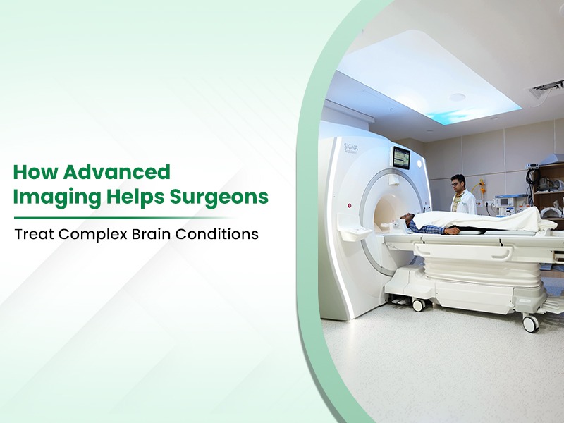
How Advanced Imaging Helps Surgeons Treat Complex Brain Conditions
In complex neurosurgical conditions, accurate imaging helps surgeons understand the brain's intricate anatomy before and during surgery. The brain contains delicate neural pathways and important blood vessels, making detailed anatomical information essential for surgical planning. Modern neurosurgery uses advanced imaging technologies to identify, analyse, and assess brain tissue and surrounding structures. These technologies can help create detailed digital maps that support surgical planning and navigation.
What Is Brain Imaging?
Brain imaging involves specialised diagnostic techniques that create detailed images of the brain and surrounding structures. Modern imaging can provide information about brain anatomy, blood flow, tissue characteristics, and, in selected cases, functional activity.
Some major components of contemporary brain imaging include:
- High-Field Magnetic Resonance: Provides detailed images of soft brain tissues, tumours, and other brain structures.
- Computed Tomography Scans: Help assess the skull, acute intracranial haemorrhage, and certain vascular or bony abnormalities, particularly in emergency settings.
- Diffusion Tensor Imaging: Maps white matter pathways and can help surgeons understand the relationship between a lesion and important neural tracts.
- Functional Brain Mapping: Helps identify areas of the brain involved in functions such as language and movement when clinically indicated.
Using these diagnostic methods allows clinical teams at Best Brain and Spine Surgery Hospital in Ghaziabad to evaluate complex neurological conditions and support appropriate treatment planning.
How Imaging Improves Surgical Planning
Before surgery, neurosurgeons use detailed imaging datasets to understand a patient's individual anatomy and plan an appropriate surgical approach. Three-dimensional reconstruction and image-guided planning can help surgeons assess the location of a lesion in relation to critical blood vessels and functional brain regions. Advanced pre-operative planning at a best brain and spine surgery hospital in Greater Noida can support careful decision-making for complex neurological procedures.
Primary advantages of pre-operative visual planning include:
- Trajectory Planning: Identifying an appropriate surgical route that avoids important brain structures where possible.
- Target Boundary Assessment: Helping distinguish a lesion from surrounding brain tissue and assess its extent.
- Vascular Mapping: Identifying important arteries and veins to support surgical planning and reduce the risk of vascular injury.
- Implant and Reconstruction Planning: Supporting the design and selection of implants or reconstruction methods for selected skull defects.
Real-Time Guidance During Surgery
Advanced imaging can also support surgeons during the procedure through intraoperative navigation and imaging systems. Surgical navigation systems use pre-operative imaging and, where appropriate, updated intraoperative information to help surgeons understand the position of surgical instruments relative to the patient's anatomy.
Key aspects of intraoperative visual guidance include:
- Real-Time Surgical Navigation: Tracking surgical instruments in relation to pre-operative imaging to support accurate localisation.
- Intraoperative Imaging: Obtaining updated images during selected procedures to assess anatomy, tissue movement, or the extent of tumour removal.
- Fluorescence-Guided Surgery: Using selected fluorescent agents that can help distinguish certain abnormal brain tumour tissue during surgery.
- Microsurgical Visualisation: High-magnification surgical microscopes help surgeons visualise delicate structures during complex procedures.
At Yashoda Medicity, advanced imaging and navigation technologies can support surgical planning and intraoperative decision-making for selected complex cranial procedures.
Benefits for Patient Safety
Integrating advanced imaging into neurosurgical care can help surgeons understand anatomy, plan surgical approaches, and reduce unnecessary disruption of important structures. The potential benefits depend on the type of condition, procedure, and individual patient factors.
Major patient safety benefits include:
- Preservation of Neurological Function: Detailed imaging can help surgeons identify important speech, motor, and other functional pathways and plan treatment accordingly.
- Targeted Surgical Access: Image-guided planning can support more targeted surgical approaches where clinically appropriate.
- Assessment of Vascular Structures: Detailed imaging can help identify important blood vessels and their relationship to the surgical target.
- Informed Surgical Decision-Making: Combining imaging findings with clinical assessment can help surgeons select an appropriate surgical strategy and anticipate anatomical challenges.
Conclusion
Advanced neuroimaging technologies provide neurosurgeons with detailed information about brain anatomy and function, supporting careful planning and image-guided treatment of complex neurological conditions. Patients seeking specialised care at a Best Brain and Spine Surgery Hospital in Noida can benefit from comprehensive diagnostic evaluation and individualised treatment planning.
At Yashoda Medicity, our neurosurgical teams use appropriate imaging, navigation, and surgical technologies to support the evaluation and treatment of complex neurological conditions. Contact us today to schedule a consultation and discuss the appropriate treatment options for your condition.
FAQs
Q1: How can high-field MRI help brain surgeons?
High-field MRI provides detailed images of brain tissue and other structures, helping neurosurgeons understand the location and characteristics of tumours or other abnormalities during treatment planning.
Q2: What are the advantages of diffusion tensor imaging during brain surgery?
Diffusion tensor imaging can map important white matter pathways and help surgeons understand their relationship to a brain lesion. This information can support surgical planning when preservation of neurological function is important.
Q3: In what way does three-dimensional surgical planning make brain surgery safer for patients?
Three-dimensional imaging and surgical planning can help surgeons understand the patient's individual anatomy and plan an appropriate route while considering important blood vessels and functional brain structures.
Q4: What is the function of real-time navigation systems in the operating theatre?
Navigation systems help surgeons track the position of surgical instruments in relation to the patient's pre-operative imaging. Accuracy varies according to the navigation system, imaging, registration, and surgical conditions, so a fixed one-millimetre accuracy should not be assumed.
Q5: How do advanced imaging methods support patient recovery after brain surgery?
Advanced imaging can support more informed surgical planning and help surgeons identify important structures. Recovery depends on the underlying condition, type of surgery, overall health, and rehabilitation needs.