 Open Emergency Modal
Open Emergency Modal
Open Emergency Modal
Overview
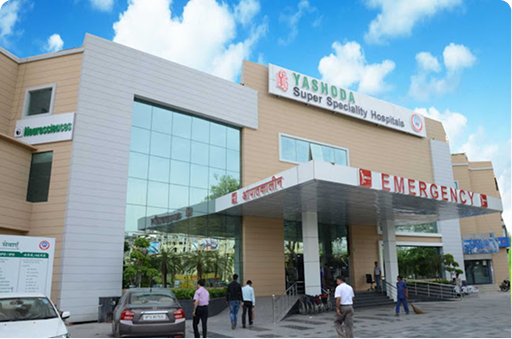
The Neonatal Intensive Care Unit at Yashoda Medicity and Yashoda Super Speciality Hospital, Kaushambi provides specialized critical care for newborn babies who are premature, low birth weight, critically ill, or born with medical complications. The NICU is equipped with advanced monitoring systems, respiratory support, thermal care facilities, and evidence-based treatment protocols to ensure safe and comprehensive care for newborns.
Our team of neonatologists, pediatric specialists, trained nurses, respiratory therapists, and support staff provides round-the-clock care for babies requiring intensive medical attention. Along with medical treatment, the department focuses on parental counseling, nutrition support, breastfeeding guidance, developmental monitoring, and family-centered care.
NICU Services Available
Conditions Treated in NICU
Technology & Infrastructure
The NICU is supported by advanced incubators, ventilators, CPAP machines, infusion pumps, neonatal resuscitation equipment, bedside ultrasonography, echocardiography, in-house laboratory, imaging support, and ECMO support for critically ill neonates.
Why Choose Our NICU
The NICU offers continuous monitoring, advanced life-support systems, specialized neonatal care, and a compassionate family-centered approach. From premature newborns to critically ill babies, the unit is designed to provide safe, timely, and expert care during the most delicate stage of life.
 More
More
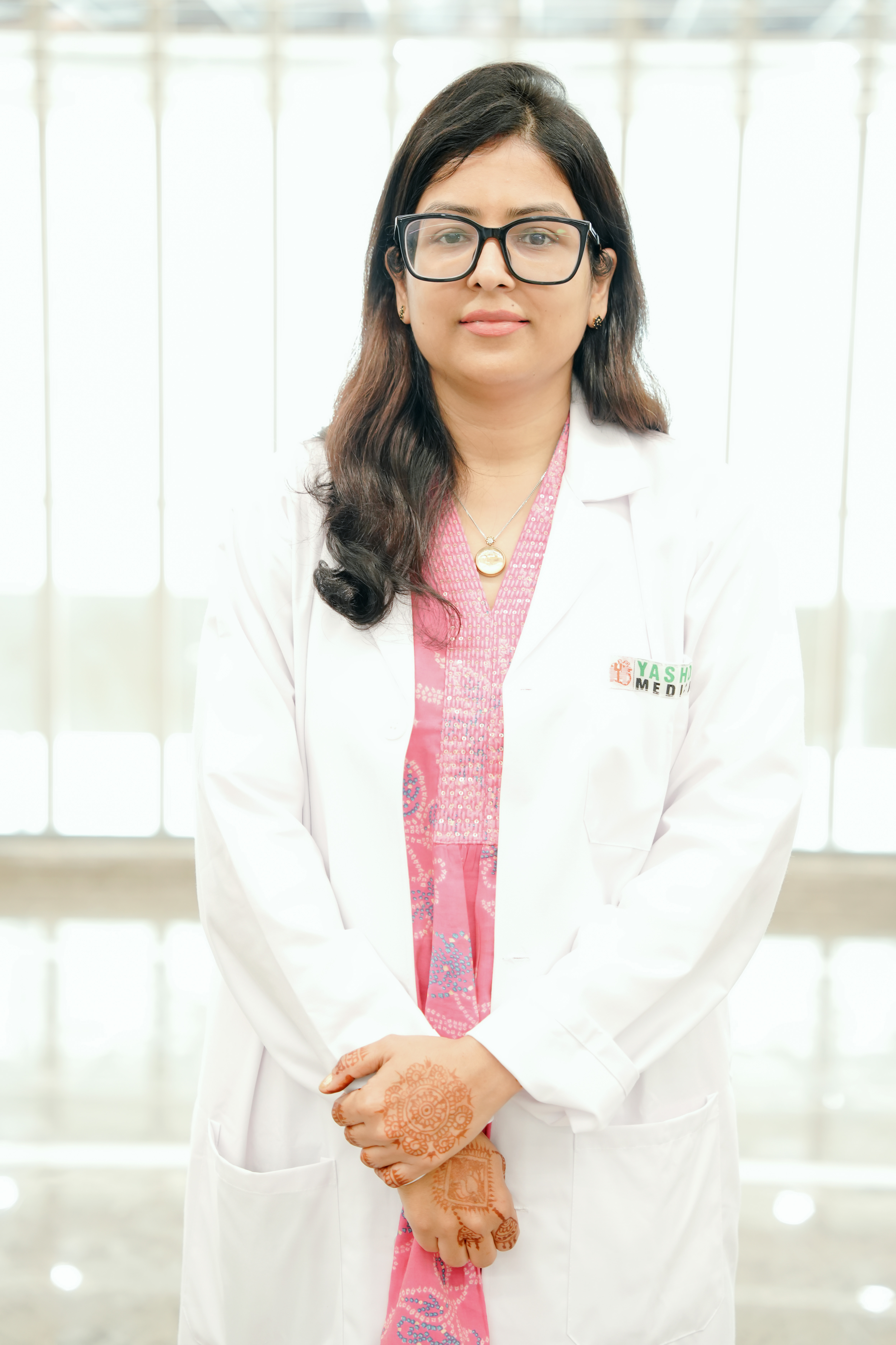
Consultant
Neonatalogist
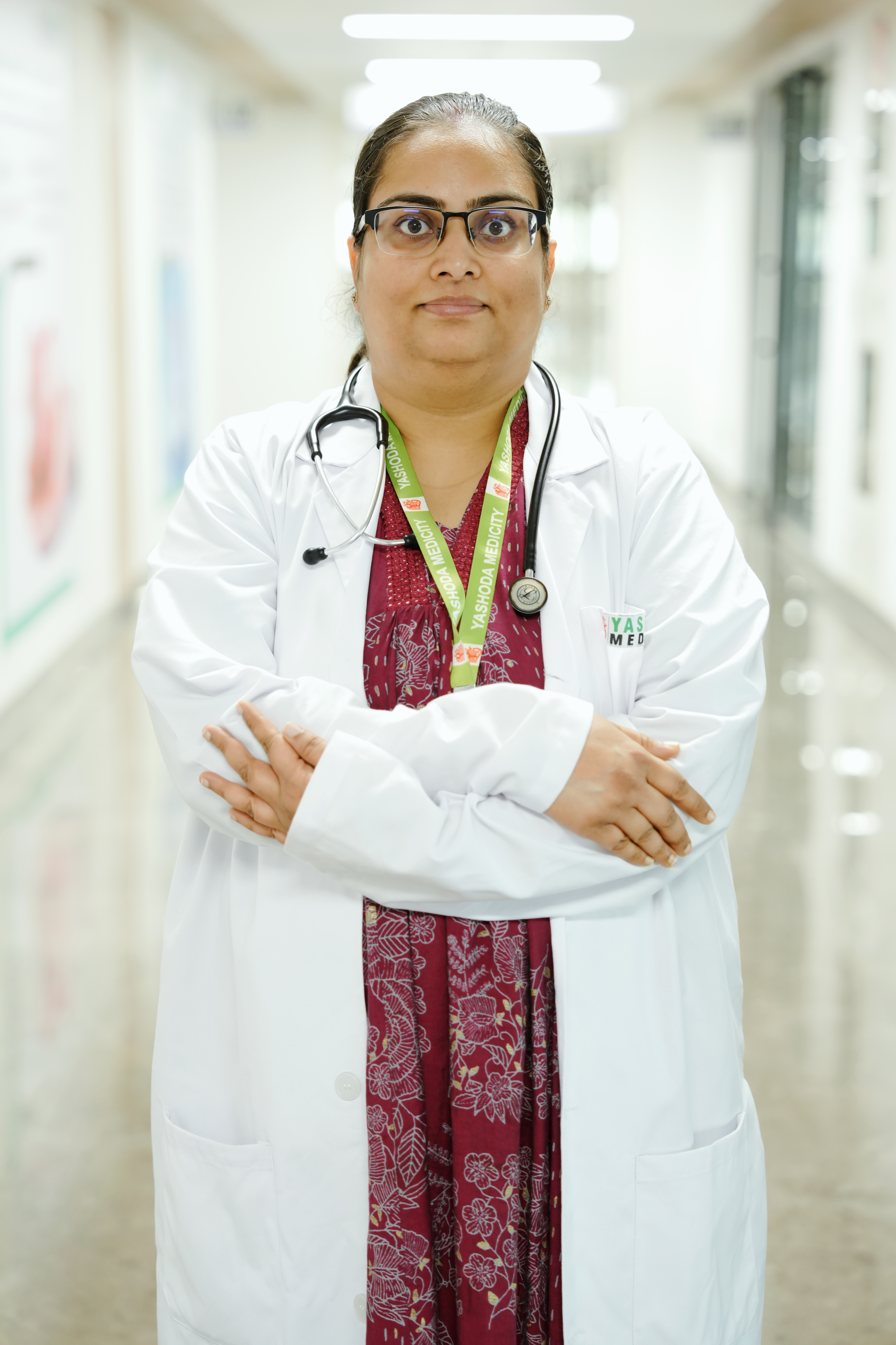
Consultant
Neonatologist
From Critical Congenital Anomalies to a Thriving Newborn: A Multidisciplinary Neonatal Triumph
The journey of a preterm infant born with multiple life-threatening congenital anomalies represents one of the greatest challenges in modern neonatal medicine. This case highlights the successful management and remarkable recovery of a late preterm baby girl born at 34 weeks of gestation. Admitted with a complex clinical presentation—including Tracheo-Esophageal Fistula (TEF), respiratory distress, a right-sided rib fracture, and a Hemodynamically Significant Patent Ductus Arteriosus (Hs-PDA)—the newborn's recovery stands as a testament to advanced neonatal expertise and seamless multidisciplinary care. Through early diagnosis, timely surgical intervention, advanced intensive care ventilation and meticulous medical management, the infant progressed from a critical condition to becoming clinically stable, feeding orally and gaining weight. Today, her recovery reflects the world-class standard of care delivered by the Neonatal Intensive Care Unit (NICU) at Yashoda Medicity.
The patient, a late preterm female infant, was delivered via Lower Segment Caesarean Section (LSCS) due to maternal polyhydramnios and leaking amniotic fluid. Although she cried immediately after birth following stimulation, routine delivery room assessment revealed an inability to pass a feeding tube into the stomach. An urgent X-ray demonstrated coiling of the feeding tube within the upper oesophageal pouch, confirming the diagnosis of Tracheo-Esophageal Fistula (TEF). This congenital condition, characterised by an abnormal connection between the oesophagus and trachea, carries an immediate risk of aspiration, respiratory failure and inability to feed.
The infant soon developed severe respiratory distress requiring immediate Continuous Positive Airway Pressure (CPAP) support and subsequently mechanical ventilation following surgery. After extubation on the second post-operative day, she developed mild tachypnoea, tachycardia and a continuous cardiac murmur. A repeat echocardiogram confirmed a large Hemodynamically Significant Patent Ductus Arteriosus (Hs-PDA) with a left-to-right shunt, placing significant strain on her cardiovascular system. Managing these multiple complex conditions required close collaboration between the departments of Neonatology, Paediatric Surgery, Paediatric Cardiology and Orthopaedics.
The cornerstone of her recovery was the successful primary surgical repair of the Tracheo-Esophageal Fistula, performed on the fourth day of life by eminent Paediatric Surgeon Dr. Minu Bajpai. Following surgery, the baby received advanced mechanical ventilation using Synchronized Intermittent Mandatory Ventilation (SIMV) with Pressure Support and Volume Guarantee (VG) mode under sedation for five days. This lung-protective ventilation strategy effectively stabilised her respiratory status, allowing safe extubation to Non-Invasive Positive Pressure Ventilation (NIPPV) and gradual weaning to room air by the eighth post-operative day.
The development of a symptomatic Hemodynamically Significant Patent Ductus Arteriosus (Hs-PDA) following extubation required prompt medical management. Simultaneously, stringent infection prevention protocols were maintained throughout the hospital stay. Broad-spectrum intravenous antibiotics were administered empirically for eight days, and the infant remained completely free from sepsis during her admission.
Through meticulous round-the-clock neonatal monitoring and comprehensive multidisciplinary care, the baby achieved an excellent recovery. Her weight increased steadily from 2.168 kg at birth to 2.260 kg at discharge, demonstrating appropriate nutritional progress. On the eighth post-operative day, a fluoroscopic oesophagogram confirmed completely normal swallowing with no evidence of an anastomotic leak, allowing gradual advancement of feeds. She successfully transitioned from orogastric tube feeding to paladai feeds on day nine, established full oral feeding by day fourteen, and was breastfeeding on demand before discharge. At the time of discharge, the infant was neurologically normal, haemodynamically stable, feeding well and had a healthy surgical wound.
A key contributor to this successful outcome was Dr. Minu Bajpai, whose exceptional surgical expertise enabled precise anatomical reconstruction of the oesophagus in this fragile newborn. Her surgical excellence laid the foundation for the infant's complete recovery and long-term well-being.
This remarkable case demonstrates the transformative impact of modern neonatal medicine, where rapid diagnosis, advanced surgical expertise, sophisticated intensive care and coordinated multidisciplinary management can successfully overcome even the most complex congenital conditions, giving vulnerable newborns the opportunity to thrive and lead healthy lives.
Treatment By:
Dr. Minu Bajpai, Dr. Srijan Singh
Overcoming the Odds: A Neonatal Breakthrough in Severe RDS and Life-Threatening PPHN
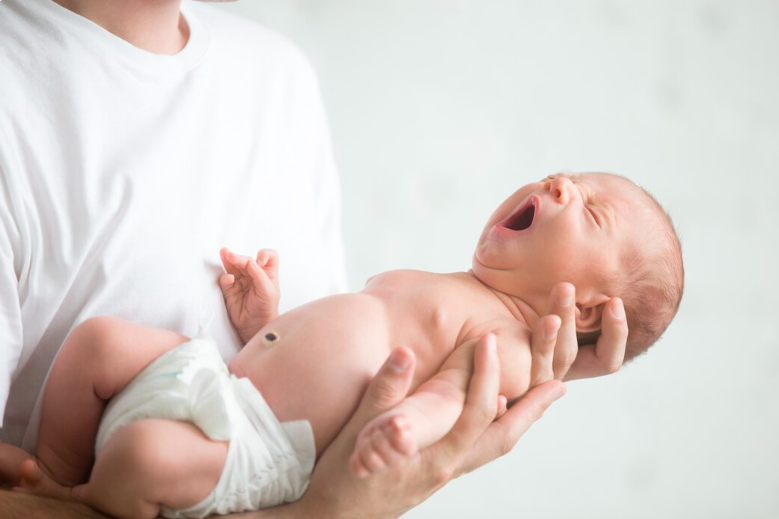
In a definitive showcase of advanced critical care capabilities, Yashoda Medicity successfully managed and treated a highly volatile clinical crisis involving an early-term neonate suffering from severe Respiratory Distress Syndrome (RDS) and Persistent Pulmonary Hypertension of the Newborn (PPHN).
The male infant was delivered via Lower Segment Caesarean Section (LSCS) at 37 weeks of gestation and was immediately transferred to the Neonatal Intensive Care Unit (NICU) after developing early signs of respiratory distress, including marked nasal flaring, tachypnoea, audible grunting and significant chest wall retractions. On admission, the baby had a Downes score of 4/10, requiring urgent Non-Invasive Positive Pressure Ventilation (NIPPV) with a high oxygen requirement. Initial chest radiographs revealed low-volume lungs, confirming Respiratory Distress Syndrome (RDS) and necessitating immediate advanced neonatal respiratory support.
The infant's condition rapidly became more complex as respiratory distress worsened despite aggressive non-invasive ventilation. The neonatal team promptly administered the first dose of Curosurf® using the advanced Less Invasive Surfactant Administration (LISA) technique, resulting in temporary stabilisation. However, approximately 12 hours later, the baby's condition deteriorated significantly, with oxygen requirements increasing to 50% along with persistent respiratory retractions.
An urgent bedside 2D echocardiography, performed by Dr. Gaurav Kumar, Paediatric Cardiologist, revealed severe pulmonary arterial hypertension (PAH) with a dilated right atrium and right ventricle, a moderate-sized Patent Ductus Arteriosus (PDA) and an aneurysmal interatrial septum. At the same time, a repeat chest X-ray demonstrated progression to Grade 3 Respiratory Distress Syndrome, indicating worsening lung disease complicated by secondary Persistent Pulmonary Hypertension of the Newborn (PPHN).
Faced with these life-threatening complications, the multidisciplinary team immediately intubated the neonate, initiated Volume-Guaranteed (VG) mechanical ventilation, provided targeted sedation and administered a second dose of surfactant.
The success of this delicate recovery was led by the meticulous clinical expertise of Dr. Srijan Singh, Consultant Neonatologist, whose precise management of neonatal haemodynamics and advanced ventilator strategies proved lifesaving. Following the second dose of surfactant, the infant showed remarkable improvement, with oxygen requirements falling to 30% and arterial blood gas parameters returning to normal.
A follow-up echocardiogram performed within 24 hours confirmed complete resolution of severe pulmonary hypertension. This significant improvement allowed successful extubation back to NIPPV, followed by gradual weaning to Continuous Positive Airway Pressure (CPAP) over the next 48 hours. The infant was subsequently transitioned off all respiratory support and maintained stable spontaneous breathing.
By the eighth day of life, the newborn had made a complete recovery from severe Respiratory Distress Syndrome (RDS) and Persistent Pulmonary Hypertension of the Newborn (PPHN). The baby demonstrated normal neurological status, steady weight gain and complete haemodynamic stability, and was discharged in excellent health.
This case highlights the exceptional standard of neonatal intensive care at Yashoda Medicity, demonstrating how early diagnosis, timely surfactant therapy, advanced ventilatory support and coordinated multidisciplinary care can successfully manage even the most complex neonatal emergencies, leading to outstanding clinical outcomes.
Treatment By:
Dr. Srijan Singh, Dr. Gaurav Kumar
Rare Anterior Urethral Diverticulum Unmasked After Posterior Urethral Valve Ablation in a Male Infant
Posterior urethral valves (PUV) represent the most common cause of congenital bladder outlet obstruction in male infants(1,2). In contrast, anterior urethral valves (AUV) and associated congenital anterior urethral diverticula (CAUD) are rare, occurring 15–30 times less frequently(3). The coexistence of both proximal (PUV) and distal (AUV/CAUD) obstructions poses unique diagnostic challenges because the dominant proximal lesion can limit distension and mask the distal anomaly until after ablation(4). Literature indicates that such combined anomalies may present with persistent or recurrent obstructive symptoms, urinary tract infections (UTIs), and upper tract deterioration post-PUV management(4–6). Early recognition via imaging (e.g., MCU) and cystoscopy is critical to prevent complications such as progressive renal damage, bladder dysfunction, or recurrent infections. This report describes a case of an anterior urethral diverticulum unmasked after PUV ablation, successfully managed with surgical reconstruction.
Case Report:
A 2-month-old male infant, born at term via normal vaginal delivery with unremarkableantenatal history, was referred as a follow-up case of PUV. On day 6 of life, he underwent cystoscopic ablation of PUV at our center for obstructive uropathy. Preoperative MCU on day2 of life demonstrated classic PUV findings . He presented at 2 months with high-grade fever (up to 102°F), poor urinary stream, dribbling, intermittent penile swelling that reduced after micturition, and dysuria. No hematuria, constipation, or neurological deficits were reported. Family history was unremarkable. On examination, the infant was irritable with mild dehydration. Abdominal palpation revealed a distended bladder. Genital examination showed ventral penile swelling that partially decompressed post-voiding . No hypospadias or other anomalies were noted. Vital signs were stable aside from fever. Labs showed leukocytosis, elevated C-reactive protein, and urine culture positive for Klebsiella pneumoniae (sensitive to targeted antibiotics). Mild azotemia was present. Ultrasonography confirmed bilateral
hydroureteronephrosis and thickened bladder wall.
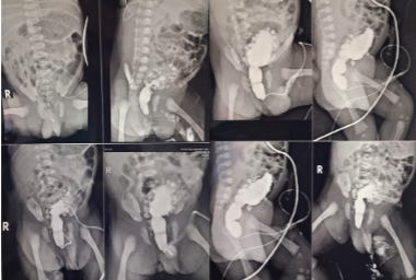
After stabilization with antibiotics and bladder drainage, a repeat MCU demonstrated a
saccular outpouching consistent with an anterior urethral diverticulum/valve, delayed
emptying, residual dilated posterior urethra, trabeculated bladder, and bilateral grade III–IV
vesicoureteral reflux (VUR) or hydroureteronephrosis . The distal
anomaly had been masked by the prior proximal obstruction.
Following resolution of infection, the infant underwent cystoscopy and urethroplasty under
general anesthesia. Intraoperative findings confirmed a significant anterior urethral
diverticulum with a valve-like membrane. The base of the diverticulum was carefully incised
to create a controlled fistula for decompression of the obstructed system . This will
be followed by meticulous urethroplasty: excision of redundant diverticular tissue,reconstruction of the urethral continuity using fine absorbable sutures to ensure a wide caliber
lumen without tension, and multilayer closure to support the repair.
The patient remained stable perioperatively. He was monitored for 24 hours with analgesia
and prophylactic antibiotics. He was discharged on postoperative day 14, afebrile, feeding
well, gaining weight, and voiding with a good stream. Follow-up ultrasonography showed
improving hydroureteronephrosis. Long-term surveillance for renal function, bladder
dynamics, and stricture is planned.
Discussion:
The association of anterior urethral diverticulum/valve with PUV is rare, with limited cases
reported(7). The proximal PUV restricts distal urethral filling, often delaying manifestation of
the anterior lesion until after ablation(4). Persistent symptoms post-PUV treatment should
prompt evaluation for concomitant distal anomalies(7).
MCU is the gold standard for diagnosis, revealing the diverticular outpouching, “spinning
top” urethra, and associated VUR or hydronephrosis(8,9). Cystoscopy confirms the valve-like
membrane and diverticulum(7).
Management is individualized based on size and tissue support. Small valves may respond to
endoscopic fulguration or laser ablation. Larger saccular diverticula, as in this case, require
open diverticulectomy and urethroplasty to excise redundant tissue, reconstruct continuity,
reduce dead space, and prevent complications (e.g., stones, recurrent infection, or
stricture)(3,10). Techniques range from primary anastomosis to staged repairs or substitution
urethroplasty in complex cases(11–13).
This case achieved excellent short-term outcomes with controlled decompression and
meticulous reconstruction. Potential complications of untreated CAUD include renal
deterioration and bladder decompensation(14,15). Thorough endoscopic evaluation and
individualized surgery are essential.
Treatment By:
Dr. Minu Bajpai, Dr. Srijan Singh
Complete Duodenal Obstruction Due to Transmesenteric Internal Bowel Herniation and Congenital Bands: A Lifesaving Neonatal Intervention
In a remarkable clinical milestone that underscores the power of swift, multidisciplinary intervention, Yashoda Medicity successfully managed and treated a highly critical, life-threatening congenital anomaly in a 4-day-old neonate. The male infant, Baby of Pushpa, was admitted to the Neonatal Intensive Care Unit (NICU) following a critical referral, presenting with an acute history of multiple episodes of bilious vomiting, significant abdominal distension, and sluggish bowel sounds. Recognizing the extreme urgency of the situation, the clinical team immediately initiated stabilization protocols, making the baby nil per os (NPO), commencing intravenous fluid resuscitation, administering empirical antibiotics, and inserting a nasogastric/orogastric tube. Initial abdominal radiographs revealed a severely dilated stomach paired with a concerning gasless abdomen, signaling a high-grade gastrointestinal obstruction that required immediate expert diagnostic elucidation and surgical evaluation.
The case rapidly transformed into a collaborative triumph through the immediate involvement of the hospital's elite pediatric medical leadership. Dr. Minu Bajpai, an eminent and veteran pediatric surgeon whose vast experience and decisive clinical acumen are widely recognized, was promptly consulted to guide the surgical trajectory. Dr. Bajpai recommended a gastrograffin contrast study, which definitively demonstrated a complete mechanical obstruction at the level of the second part of the duodenum alongside proximal gastro-duodenal dilatation. Armed with this critical insight, Dr. Bajpai led the surgical team in performing an emergent exploratory laparotomy. The intraoperative findings revealed a rare and treacherous internal internal herniation of the small bowel through the transverse mesocolon, compounded by restrictive congenital bands compromising the second part of the duodenum. With meticulous precision, the surgical team divided the bands and successfully achieved complete deherniation of the entrapped bowel, resolving the primary anatomical crisis.
Following the intricate surgical procedure, the complex task of critical post-operative management fell to the highly accomplished neonatologist, Dr. Srijan Singh, whose specialized dedication to neonatal intensive care proved pivotal to the infant’s survival. Dr. Singh orchestrated a comprehensive, advanced recovery plan in the NICU, keeping the fragile neonate NPO and introducing Total Parenteral Nutrition (TPN) via a peripherally inserted central catheter (PICC) line to sustain metabolic demands. Under Dr. Singh’s vigilant oversight, the baby received tailored mechanical ventilation for 36 hours before being successfully extubated to room air. Dr. Singh ordered a repeat gastrograffin study, which confirmed a perfectly patent, non-obstructed post-operative duodenum with excellent contrast passage.
The synergistic collaboration between the surgical prowess of Dr. Minu Bajpai and the meticulous, expert neonatal care of Dr. Srijan Singh ensured a flawless trajectory toward recovery. Over the subsequent days, enteral feeds were gradually advanced, the post-operative sepsis screen remained entirely negative, and blood cultures proved sterile, allowing the safe discontinuation of intravenous antibiotics. By the tenth post-operative day, the infant demonstrated a complete clinical turnaround—actively accepting full breastfeeds and paladai feeds, passing normal stools without any vomiting, and steadily gaining weight. The infant was discharged in a hemodynamically stable condition with a healthy, healing surgical site. This extraordinary breakthrough highlights the world-class neonatal and pediatric surgical capabilities available at Yashoda Medicity, showcasing how seamless teamwork, clinical expertise, and cutting-edge intensive care can rewrite the medical outcomes for the most vulnerable patients.
Treatment By:
Dr. Minu Bajpai, Dr. Srijan Singh
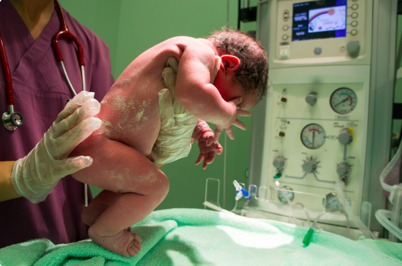
A primigravida mother with pregnancy-induced hypertension (PIH) and deranged antenatal Dopplers (reversal of flow in ductus venosus) delivered baby via emergency LSCS at 30 weeks. Baby had extremely low birth weight (900 grams) and had been diagnosed with severe fetal growth restriction (FGR) antenatally and presented with perinatal depression and respiratory distress immediately after birth.
Neonatal Course & Challenges: This extremely preterm, extremely low birth weight (ELBW) infant faced several serious complications typical of extreme prematurity:
Despite these multiple challenges, the baby showed remarkable resilience.
Multidisciplinary Management: Under the leadership of Dr. Srijan Singh, the NICU team at Yashoda Medicity provided comprehensive, evidence-based care:
The baby remained hemodynamically stable in the later part of the stay, achieved full oral (paladai) feeds, and maintained temperature and vitals without support.Baby was neurologically normal at discharge and gaining weight. Baby did not have any ROP, BPD or IVH.
Treatment By:
Dr. Srijan Singh, Dr. Samreen Zehra
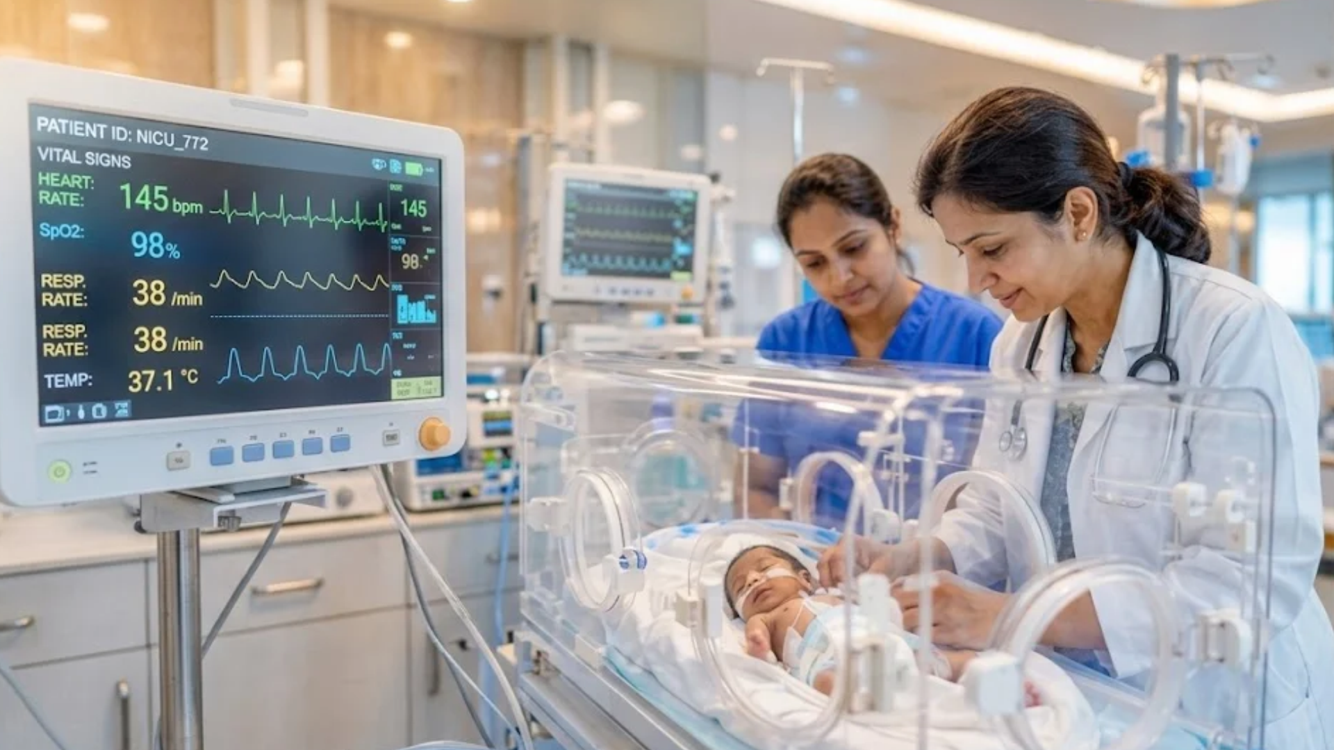
Advanced neonatal care plays a critical role in improving survival and long-term outcomes for extremely preterm infants. In a recent case, our multidisciplinary team successfully managed the delivery and care of a baby boy born at just 26 weeks of gestation, weighing under 800 grams.
The newborn required immediate respiratory support and was admitted to a Level III Neonatal Intensive Care Unit (NICU), where he received round-the-clock monitoring and specialised care. Through meticulous medical management and dedicated nursing support, the baby demonstrated consistent progress over the course of treatment.
He was discharged in a stable condition, breathing independently and feeding well, marking a significant milestone in his recovery journey.
This case was led by Dr. Neha Agrawal, Consultant – Pediatrics & Neonatology; Dr. J.B. Sharma, Principal Director & Head – Obstetrics and Gynaecology; Dr. Namita Nigam Datta, Senior Consultant – Obstetrics and Gynaecology; and Dr. Nisha, Consultant – Obstetrics & Gynaecology, alongside our experienced nursing and clinical teams.
It reflects the strength of coordinated clinical expertise, advanced neonatal infrastructure, and a commitment to delivering specialised care focused on giving every newborn the best possible start in life.
Treatment By:
Dr. Neha Agrawal, Dr. J.B. Sharma, Dr. Namita Nigam Datta, Dr. Nisha

A G6A5L0 mother with an IVF-conceived pregnancy had a history of threatened abortion and intrauterine demise of Twin A at 22 weeks of gestation.
Twin B, a 24-week gestation male/female preterm neonate weighing 650 g, was delivered by emergency LSCS in view of placental abruption. The baby cried immediately after birth but developed severe respiratory distress and was promptly shifted to the Level III NICU for intensive care.
The neonate received three doses of surfactant therapy and required invasive mechanical ventilation for 7 days, followed by non-invasive respiratory support, with gradual weaning to room air. Enteral feeding was initially limited due to feed intolerance, and the baby remained on total parenteral nutrition (TPN) until full enteral feeds were successfully established by Day 18 of life.
During the NICU stay, the baby was treated with intravenous antibiotics for 2 weeks for sepsis. Subsequently, the baby developed fungal sepsis, which was successfully managed with appropriate antifungal therapy.
Serial cranial ultrasonography was performed as per protocol and showed no evidence of intraventricular hemorrhage (IVH) or periventricular leukomalacia (PVL).
ROP screening was initially normal. However, at 33 weeks postmenstrual age, the baby developed pre-plus disease in Zone II, which required laser photocoagulation therapy. Subsequent follow-up examinations documented satisfactory regression of ROP.
The baby also required packed red blood cell (PRBC) transfusion during the hospital stay for anemia of prematurity.
The case was successfully managed by a multidisciplinary team comprising Dr. Minu Bajpai, Dr. J.B. Sharma, Dr. Neha, Dr. Samreen, Dr. Divya Arora, Dr. Ayush Jain, and Dr. Nisha.
After a prolonged NICU stay of 78 days, the baby was discharged in a clinically stable condition on room air, tolerating spoon feeds and breastfeeding well, with a discharge weight of 1,625 g.
Treatment By:
Dr Neha Agrawal (Consultant Neonatologist ) and NICU team.
It was a very good experience, I had a cholecystectomy. All the doctors including surgeon, anesthetic, nursing and housekeeping staff were very supportive. I was admitted for 24 hours. Quick and very fast recovery. Under Dr P.K.Dewan, sir is very humble and nice.
All experience good, patient treatment was good, satisfied with all the services, nursing staff and doctors team also good and cooperative.
I had an excellent experience at Yashoda Hospital for my mother’s Total Knee Replacement Surgery.
From a clinical perspective, Dr. Amit Sharma and his team were outstanding. Their expertise and professionalism were evident throughout the process, and they did a fantastic job with the surgery and post-operative care.
Beyond the medical treatment, the administrative side was equally impressive. Mr. Varun took complete charge of the Insurance (TPA) and admission process, ensuring everything was seamless and stress-free for our family. His coordination made a significant difference in our overall experience.
We are truly grateful to the entire team for their dedication and care. Highly recommended!
Mr. Ajeet Kumar Verma from Yashoda Homecare provided satisfactory service of sample collection. Well behaved, humble & polite person
I had really good experience with Yashoda Home Care, the sample collection was very hygienically taken and pain-free, hassle-free. Mr. Ajeet was the phlebotomist.
Had a good experience with Ajeet Kumar Verma from Yashoda Home Care who supported me while I was looking for someone to dress my mom’s stitches. He is very professional and a caring person.
Great support and service by the Home care department. Everyone from Ms Niharika, Mr. Anoop to all the attendants including Mr. Natwar Pandey, Mr Inam and Mr. Ajit, all have been superbly supportive and helpful throughout. God bless you all and highly recommended.
Very good services in home care Yashoda hospital his ish
Caretaker Deepanshu and Jatin and Satish is very good caretaker I have never seen better care takers in my life.
And he is very polite behaviour and friend in nature
Yashoda Super Specialty Hospitals Kaushambi service is very good according other hospital and other home care services
The home care facility provided by the hospital is also good, the staff which was at home was cooperative.
Home care services are Good. Mr. सुनील Sajwan was cooperative and the केयर taker Mrs. Shushila's वर्क has been quite satisfactory.
Had a. Very good experience with Yashoda Homecare, Kaushambi. The staff was very cooperative with excellent on time services. I would like to give a special mention to Mr Sunil Sajwan (Sales) and Sushila ji (PCA) for their quick response and excellent services.
Fully satisfied Mr .Ajeet Kumar Verma service Yashoda home care . vry good
Was great to recieve the best homecare services from yashoda hospital and really impressed with Ms. Niharika's support at the initial phase followed by Mr. Natwar Pandey who was really supportive in taking care of my father basic daily needs.
Thanks for all the support


 Call: +91 9266610101